MUCORMYCOSIS - MY NOTES PERIODONTOLOGY PART II
Scene 1 (0s)
THUMBNAIL 2. Fumident logo small. MY NOTES PERIODONTOLOGY.
Scene 2 (7s)
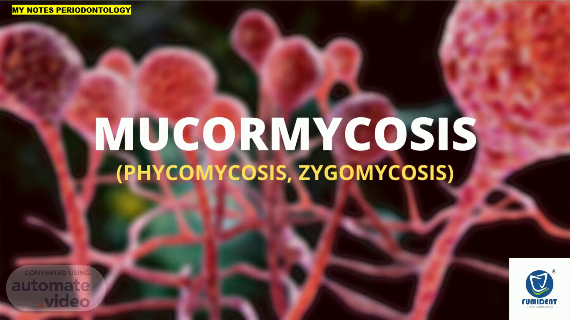
22. MUCORMYCOSIS (PHYCOMYCOSIS, ZYGOMYCOSIS) ( FROM PART II - SECTION 2: IMAGES & IDENTIFICATION) By Dr. Manish A. Ashtankar BDS, MDS, PhD Scholar Author of book MY NOTES PERIODONTOLOGY Part I, II, III.
Scene 3 (14s)
Screenshot (770). 1. Fumident logo small. MY NOTES PERIODONTOLOGY.
Scene 4 (20s)
Screenshot (771). Fumident logo small. MY NOTES PERIODONTOLOGY.
Scene 5 (27s)
Screenshot (772). Fumident logo small. MY NOTES PERIODONTOLOGY.
Scene 6 (34s)
DISCLAIMER. This video is for BDS, MDS student for last minute revision on different topics which may hep them in theory, practical, entrance exams. This video can help general practioner to improve their knowledge on different topics which may help them to reach dignosis correctly. Information in this video is taken from different sources like books, article whose references are given at the end. We further request viewers to confirm it. In case of any dispute because of information given here, we will not be responsible..
Scene 7 (39s)
WHAT IS MUCORMYCOSIS?. Mucormycosis is a rare opportunistic fungal infection, commonly seen in patients with immunocompromised conditions such as uncontrolled diabetes mellitus and leukaemias and COVID-19. What is the Phycomycosis and Zygomycosis? Phycomycosis is an uncommon condition of the gastrointestinal tract and skin most commonly found in dogs and horses. The condition is caused by a variety of molds and fungi, and individual forms include pythiosis, zygomycosis, and lagenidiosis. Pythiosis is the most common type and is caused by Pythium, a type of water mould. Zygomycosis can also be caused by two types of zygomycetes, Entomophthorales (such as Basidiobolus and Conidiobolus) and Mucorales (such as Mucor, Mortierella, Absidia, Rhizopus, Rhizomucor, and Saksenaea). The latter type of zygomycosis is also referred to as mucormycosis . (source wikipedia).
Scene 8 (1m 13s)
CLINICAL FORMS. Fumident logo small. MY NOTES PERIODONTOLOGY.
Scene 9 (1m 34s)
Fig. 1: Vestibular mucormycosis and aspergillosis: the vestibular ulcerative lesion in the area of the teeth 25-27.
Scene 10 (1m 55s)
SPREAD. Found in fruits, soil, dust, and manure. The infections spread from inhalation of spores through the nose or mouth, or sometimes through a skin laceration. Mucorales are ubiquitous moulds, profusely recovered from the decaying organic matter. Evidences from studies have revealed heavy mould spore counts in hospital air due to hot, humid environment in our tropical climate. It can be cultured from the oral cavity, nasal passages, throat and stools of healthy patients without a sign of disease..
Scene 11 (2m 20s)
SYMPTOMS. Symptoms of rhinocerebral (sinus and brain) mucormycosis include: One-sided facial swelling Headache Nasal or sinus congestion Black lesions on nasal bridge or upper inside of mouth that quickly become more severe Fever Symptoms of pulmonary (lung) mucormycosis include: Fever Cough Chest pain Shortness of breath.
Scene 12 (2m 37s)
Cutaneous (skin) mucormycosis can look like blisters or ulcers, and the infected area may turn black. Other symptoms include pain, warmth, excessive redness, or swelling around a wound. Symptoms of gastrointestinal mucormycosis include: Abdominal pain Nausea and vomiting Gastrointestinal bleeding. Disseminated mucormycosis typically occurs in people who are already sick from other medical conditions, so it can be difficult to know which symptoms are related to mucormycosis. Patients with disseminated infection in the brain can develop mental status changes or coma. References Petrikkos G, Skiada A, Lortholary O, Roilides E, Walsh TJ, Kontoyiannis DP. Epidemiology and clinical manifestations of mucormycosisexternal icon. Clin Infect Dis. 2012 Feb;54 Suppl 1:S23-34. Lewis RE, Kontoyiannis DP. Epidemiology and treatment of mucormycosisexternal icon. Future Microbiol. 2013 Sep;8(9):1163-75. Spellberg B, Edwards Jr. J, Ibrahim A. Novel perspectives on mucormycosis: pathophysiology, presentation, and managementexternal icon. Clin Microbiol Rev. 2005 Jul;18(3):556-69. Ribes JA, Vanover-Sams CL, Baker DJ. Zygomycetes in human diseaseexternal icon. Clin Microbiol Rev 2000; 13:236-301..
Scene 13 (3m 26s)
CLINICAL FEATURES. Malaise, headache, facial pain, swelling and low grade fever 5 . The disease initiates from nasal mucosa or palate and extends to the paranasal sinuses spreading through the surrounding vessels. Can involve the retro-orbital region by direct extension 4 . Proptosis, ptosis, pupillary dilatation, orbital cellulitis and loss of vision can occur with loss of function of cranial nerve III, IV, VI with orbital involvement 5 . In mucormycosis , the fungal hyphae start to attack local tissue. Direct penetration and growth through the wall of blood vessels are responsible for the propensity for thrombosis and tissue necrosis..
Scene 14 (3m 56s)
RADIOLOGICAL PICTURE. Shows opacification of the paranasal sinuses without fluid level, thickening of the sinus mucosa and bone distraction of the sinus walls..
Scene 15 (4m 12s)
DIAGNOSIS. From tissue biopsy, clinical picture Histological picture : are characterised by extensive tissue necrosis and the presence of numerous, large (around 5–30 μm). These fungal hyphae are thin-walled, non-septate, branched at right angles and have a ribbon-like appearance..
Scene 16 (4m 40s)
MANAGEMENT. Medical and surgical intervention. Treatment of underlying medical condition. Systemic antifungal agents: Amphotericin B, Liposomal Amphotericine B. Surgical: debridement of all infected and necrotic tissues. Radical resection, partial or total maxillectomy, mandibulectomy and orbital operation are required in some cases..
Scene 17 (4m 58s)
PROGNOSIS. In most cases, the prognosis is poor, mortality rates depending on its form and severity. In the rhinocerebral form, the mortality rate : 30% to 70%, Disseminated mucormycosis : up to 90%, Patients with AIDS : almost 100%. Possible complications include the partial loss of neurological function, blindness and clotting of brain or lung vessels..
Scene 18 (5m 17s)
REFERENCES. 1.deShazo RD, O’Brien M, Chapin K, Soto-Aguilar M, Gardner L, Swain R. A new classification and diagnostic criteria for invasive fungal sinusitis. Arch Otolaryngol Head Neck Surg. 1997;123:1181– 1188. 2.Tryfon S, Stanopoulos I, Kakavelas E, Nikolaidous A, Kioumis I. Rhinocerebral Mucormycosis in a Patient with latent diabetes mellitus: A Case Report. J Oral Maxillofac Surg. 2002;60:328– 330. 3.Peterson KL, Wang M, Canalis RF. Rhinocerebral Mucormycosis: Evaluation of the Disease and Treatment Options. Laryngoscope. 1997;107:855– 861. 4.Sugar AM. Mucormycosis. Clin Infect Dis. 1992;14:126–129. 5.Taylor R, Shlkar G, Budson R, et al: Mucormycosis of the oral mucosa. Arch Dermatol 89:419, 1964. 6.Ribeiro NNF, Cousin GCS, Wilson GE, Butterworth DM, Woodwards RTM. Lethal invasive mucormycosis: Case report and recommendation for treatment. Int J Oral Maxillofac Surg.2001;30:156–159. 7.Rudramurthy SM, Singh G, Hallur V et al. High fungal spore burden with predominance of Aspergillus in hospital air of a tertiary care hospital in Chandigarh. Indian J Med Microbiol2016; 34:529-532..
Scene 19 (6m 9s)
MY NOTE 127 and a rare 2. The The : . fig are d Radu.
Scene 20 (6m 18s)
MY NOTE rue WTES 3: NSEASZ. Fumident logo small. MY NOTES PERIODONTOLOGY.
Scene 21 (6m 25s)
1. 1. 1. Fumident logo small.
Scene 22 (6m 32s)
HOW TO GET THE BOOKS? FOR PRE-BOOKING WHATS APP ON 9284203288.
Scene 23 (6m 59s)
MY NOTES PERIODONTOLOGY PART 2 ABOUT AUTHORS DS ECCH v-y.ws- •S..AÄQ•«" BOS. N" v-Y-WS- Anva•atL THANK you FOR WATCHING.