Scene 1 (0s)
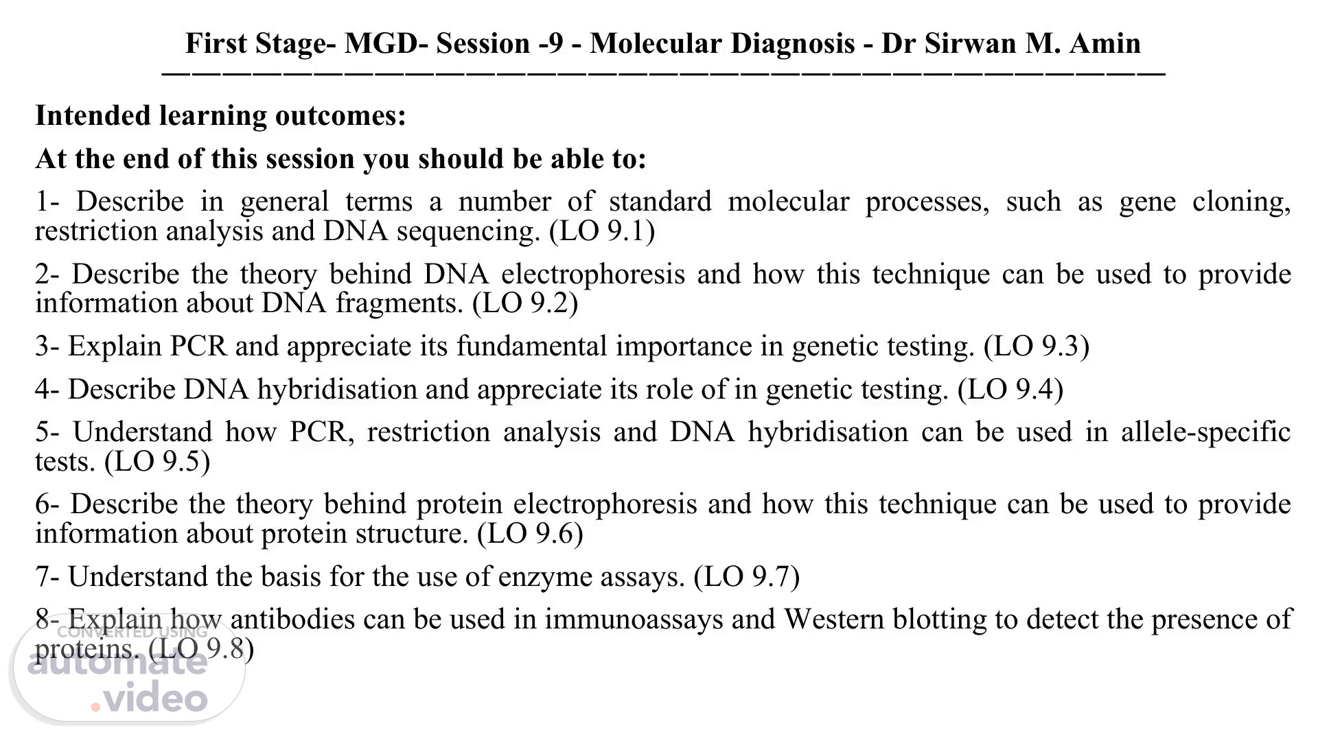
[Audio] First Stage- MGD- Session -9 - Molecular Diagnosis - Dr Sirwan M. Amin ――――――――――――――――――――――――――――――――― Intended learning outcomes: At the end of this session you should be able to: 1- Describe in general terms a number of standard molecular processes, such as gene cloning, restriction analysis and DNA sequencing. (LO 9.1) 2- Describe the theory behind DNA electrophoresis and how this technique can be used to provide information about DNA fragments. (LO 9.2) 3- Explain PCR and appreciate its fundamental importance in genetic testing. (LO 9.3) 4- Describe DNA hybridisation and appreciate its role of in genetic testing. (LO 9.4) 5- Understand how PCR, restriction analysis and DNA hybridisation can be used in allele-specific tests. (LO 9.5) 6- Describe the theory behind protein electrophoresis and how this technique can be used to provide information about protein structure. (LO 9.6) 7- Understand the basis for the use of enzyme assays. (LO 9.7) 8- Explain how antibodies can be used in immunoassays and Western blotting to detect the presence of proteins. (LO 9.8).
Scene 2 (1m 30s)
[Audio] First Stage- MGD- Session -9 - Molecular Diagnosis ――――――――――――――――――――――――――――――――― ANALYSIS OF PROTEINS ANALYSIS OF DNA 1. Analysis of DNA - at the molecular level 1. Analysis of proteins - Gel electrophoresis - DNA sequencing a) SDS-PAGE b) Isoelectric focusing 2. Analysis of DNA - at the gene level c) Two-dimensional electrophoresis (2D-PAGE) a) Restriction analysis / Gene cloning b) Gel electrophoresis 2. Analysis of proteins - Immunological techniques c) Southern blotting / hybridisation a) Western blotting d) Polymerase Chain Reaction (PCR) b) Enzyme-linked immunoabsorbent assays (ELISA) 3. Analysis of DNA - at the chromosome level 3. Analysis of proteins - Enzyme assays a) Karyotyping b) Fluorescent in situ hybridisation (FISH).
Scene 3 (2m 47s)
[Audio] First Stage- MGD- Session -9 - Molecular Diagnosis ――――――――――――――――――――――――――――――――― Why do we need to know all this? Advances in modern molecular genetics have enabled the development of a wide range of approaches for the identification and analysis of genes and specific alleles. To understand these approaches and their use in tracking genes through families it is important to comprehend some of the basic molecular techniques that are used in analysis of DNA, such as gene cloning, restriction analysis, DNA sequencing, DNA hybridisation (Southern blotting) and Polymerase Chain Reaction (PCR). This is important for helping to diagnose disease and to monitor the progression of the disease and its treatment. Although many different biological molecules can be analysed, we will focus on techniques used to analyse proteins, such as protein gel electrophoresis and enzyme assays..
Scene 4 (3m 48s)
[Audio] First Stage- MGD- Session -9 - Molecular Diagnosis ――――――――――――――――――――――――――――――――― ANALYSIS OF DNA 1. Analysis of DNA - at the molecular level DNA sequencing The ultimate analytical assay for DNA has to be the determination of the order in which the individual nucleotides are linked together to form the DNA molecule. The technique to do so is called DNA sequencing. Discovered in the early 1970s, and established fully in 1975 with the Sanger dideoxy chain termination method, DNA sequencing has now become a fully automated process. Automation of this technique was crucial for genomics, the study of entire genomes of organisms. The 3000 million nucleotides that make up the human genome were fully sequenced by 2000, and the first full assembly was published in 2003..
Scene 5 (4m 48s)
[Audio] First Stage- MGD- Session -9 - Molecular Diagnosis ――――――――――――――――――――――――――――――――― 1. Analysis of DNA - at the molecular level DNA sequencing Sanger dideoxy chain termination method.
Scene 6 (5m 2s)
[Audio] First Stage- MGD- Session -9 - Molecular Diagnosis ――――――――――――――――――――――――――――――――― ANALYSIS OF DNA 2. Analysis of DNA - at the gene level a) Restriction analysis / Gene cloning Restriction endonucleases (restriction enzymes) are bacterial enzymes that are able recognise specific DNA sequences (restriction site) and then cut the double-stranded DNA at that specific site. Restriction sites are often only a few nucleotides long (most often 4 or 6 bp, occasionally 8 or more bp). The double stranded break in the DNA molecule can be repaired with the enzyme DNA ligase. Restriction enzymes are crucial tools in many molecular biological techniques, such as gene cloning..
Scene 7 (6m 4s)
[Audio] First Stage- MGD- Session -9 - Molecular Diagnosis ――――――――――――――――――――――――――――――――― ANALYSIS OF DNA 2. Analysis of DNA - at the gene level a) Restriction analysis / Gene cloning.
Scene 8 (6m 21s)
[Audio] What is DNA ligase? Joined by DNA ligase DNA ligase is a specific type of enzyme, a ligase, that facilitates the joining of DNA strands together by catalyzing the formation of a phosphodiester bond.
Scene 9 (6m 39s)
[Audio] First Stage- MGD- Session -9 - Molecular Diagnosis ――――――――――――――――――――――――――――――――― ANALYSIS OF DNA 2. Analysis of DNA - at the gene level b) Gel electrophoresis As DNA molecules are negatively charged, they will move towards the positively electrode when placed in an electric field. When DNA molecules are placed in a gel in an electric field, smaller DNA molecules will be able to travel faster than larger DNA fragments. This forms the basic principle of DNA gel electrophoresis. DNA fragments can be separated on size using this technique. After electrophoresis DNA can be visualised under UV light, when ethidium bromide, a chemical that binds to the DNA and fluoresces under UV light, is added during the process..
Scene 10 (7m 33s)
[Audio] First Stage- MGD- Session -9 - Molecular Diagnosis ――――――――――――――――――――――――――――――――― ANALYSIS OF DNA 2. Analysis of DNA - at the gene level b) Gel electrophoresis.
Scene 11 (7m 50s)
[Audio] First Stage- MGD- Session -9 - Molecular Diagnosis ――――――――――――――――――――――――――――――――― ANALYSIS OF DNA 2. Analysis of DNA - at the gene level c) Southern blotting / hybridisation Following DNA gel electrophoresis the separated DNA molecules can be transferred to a membrane and specific DNA fragments of interest can be visualised using a labelled probe. The process whereby DNA is transferred from the gel to a solid membrane is referred to as 'blotting'. Hybridisation is the process whereby two single-stranded complementary DNA sequences reform hydrogen bonds between the complementary bases, and in doing so form a double-stranded DNA fragment. In Southern hybridisation, a single-stranded DNA molecule labelled with radioactive or fluorescent tags, is used as probe, and will be able to 'find' and hybridise with any complementary DNA sequences present on the membrane. (NOTE: If RNA instead of DNA is analysed this technique is referred to as Northern blotting.).
Scene 12 (9m 0s)
[Audio] First Stage- MGD- Session -9 - Molecular Diagnosis ――――――――――――――――――――――――――――――――― 2. Analysis of DNA - at the gene level c) Southern blotting / hybridisation What is Probe? A probe is a single-stranded sequence of DNA or RNA used to search for its complementary sequence in a sample genome..
Scene 13 (9m 27s)
[Audio] First Stage- MGD- Session -9 - Molecular Diagnosis ――――――――――――――――――――――――――――――――― ANALYSIS OF DNA 2. Analysis of DNA - at the gene level d) Polymerase Chain Reaction (PCR) By far the most powerful technique to analyse DNA is called Polymerase Chain Reaction or PCR. The technique is based on repeated cycles of denaturation, hybridisation and DNA synthesis. In this way thousands of millions copies of a specific DNA fragment can be created from one DNA template molecule. In PCR is, primers must bind specifically to a known sequence on the template DNA. However, clever use of PCR means that this powerful and very sensitive technique can also be used to discover many novel sequences. One is the biggest strengths of PCR is its sensitivity which means that the template DNA amount can be minute and could even come from a very crude sample like a mouth swap. PCR is the technique of choice for the diagnosis of many inherited diseases; it has also been used to detect the presence of tumour cells and the very early stages of infection by pathogenic microorganisms or viruses..
Scene 14 (10m 52s)
[Audio] First Stage- MGD- Session -9 - Molecular Diagnosis ――――――――――――――――――――――――――――――――― ANALYSIS OF DNA 2. Analysis of DNA - at the gene level d) Polymerase Chain Reaction (PCR).
Scene 15 (11m 7s)
[Audio] First Stage- MGD- Session -9 - Molecular Diagnosis ――――――――――――――――――――――――――――――――― ANALYSIS OF DNA 3. Analysis of DNA - at the chromosome level a) Karyotyping A karyotype is a picture of the full set of stained metaphase chromosomes of an individual organised according to chromosome number..
Scene 16 (11m 32s)
[Audio] First Stage- MGD- Session -9 - Molecular Diagnosis ――――――――――――――――――――――――――――――――― ANALYSIS OF DNA 3. Analysis of DNA - at the chromosome level b) Fluorescent in situ hybridisation (FISH) This technique allows the investigation of specific DNA sequences on chromosomes inside the cell. Fluorescent probes for a specific gene (or several genes) can be used or probes for specific DNA stretches on chromosomes, like telomeres or centromeres. For chromosome investigation chromosome painting is often used, whereby each chromosome is visualised using a different coloured fluorescent probe.
Scene 17 (12m 19s)
[Audio] First Stage- MGD- Session -9 - Molecular Diagnosis ――――――――――――――――――――――――――――――――― ANALYSIS OF PROTEINS 1. Analysis of proteins - Gel electrophoresis Because proteins are usually charged, this means that if they are placed in an electric field then they will move towards the relevant electrode. Positively charged (cationic or basic) proteins will move towards the negative electrode (cathode), whilst negatively charged (anionic or acidic) proteins will move towards the positively electrode (anode). The migration of native folded proteins in a gel is dependent not only on their charge but also the size and shape of the molecule..
Scene 18 (13m 9s)
[Audio] First Stage- MGD- Session -9 - Molecular Diagnosis ――――――――――――――――――――――――――――――――― ANALYSIS OF PROTEINS 1. Analysis of proteins - Gel electrophoresis a) SDS-PAGE Sodium dodecyl sulphate polyacrylamide gel electrophoresis (SDS-PAGE) is a technique which allows proteins to be separated purely on the basis of their molecular weight. The detergent SDS denatures protein molecules and one molecule of SDS binds for every 2 amino acids. The bound SDS has a large negative charge which masks the intrinsic charge of the protein. Therefore, proteins are separated purely on the basis of molecular weight. After electrophoresis proteins can be visualised by adding a dye, such as Coomassie Blue, which binds to the proteins but not to the gel..
Scene 19 (14m 11s)
[Audio] First Stage- MGD- Session -9 - Molecular Diagnosis ――――――――――――――――――――――――――――――――― ANALYSIS OF PROTEINS 1. Analysis of proteins - Gel electrophoresis a) SDS-PAGE.
Scene 20 (14m 26s)
First Stage- MGD- Session -9 - Molecular Diagnosis ――――――――――――――――――――――――――――――――― ANALYSIS OF PROTEINS 1. Analysis of proteins - Gel electrophoresis a) SDS-PAGE.
Scene 21 (14m 31s)
[Audio] First Stage- MGD- Session -9 - Molecular Diagnosis ――――――――――――――――――――――――――――――――― ANALYSIS OF PROTEINS 1. Analysis of proteins - Gel electrophoresis b) Isoelectric focusing Proteins can also be separated on the basis of the isoelectric points (pI). In this case native proteins are applied to a gel containing a pH gradient. A protein will migrate in the electric field until it reaches a pH that matches its pI–at this point the protein will have no overall net charge and so will stop migrating..
Scene 22 (15m 8s)
[Audio] First Stage- MGD- Session -9 - Molecular Diagnosis ――――――――――――――――――――――――――――――――― ANALYSIS OF PROTEINS 1. Analysis of proteins - Gel electrophoresis b) Isoelectric focusing.
Scene 23 (15m 17s)
[Audio] First Stage- MGD- Session -9 - Molecular Diagnosis ――――――――――――――――――――――――――――――――― ANALYSIS OF PROTEINS 1. Analysis of proteins - Gel electrophoresis c) Two-dimensional electrophoresis (2D-PAGE) This technique combines isoelectric focusing and SDS-PAGE to separate proteins in complex mixtures. 2D-PAGE allows the separation of proteins that have identical pI values but different molecular weights, or proteins of identical pI that differ in molecular weight. This is an important technique for proteomics..
Scene 24 (16m 3s)
[Audio] First Stage- MGD- Session -9 - Molecular Diagnosis ――――――――――――――――――――――――――――――――― ANALYSIS OF PROTEINS 2. Analysis of proteins - Immunological techniques Antibodies can be generated that are highly specific for a particular protein. This specificity is important as it allows for the identification of particular proteins in solution. a) Western blotting Following SDS-PAGE the separated proteins can be transferred to a membrane and specific proteins can be visualised using antibody binding..
Scene 25 (16m 41s)
[Audio] First Stage- MGD- Session -9 - Molecular Diagnosis ――――――――――――――――――――――――――――――――― ANALYSIS OF PROTEINS 2. Analysis of proteins - Immunological techniques a) Western blotting Following SDS-PAGE the separated proteins can be transferred to a membrane and specific proteins can be visualised using antibody binding..
Scene 26 (17m 5s)
[Audio] First Stage- MGD- Session -9 - Molecular Diagnosis ――――――――――――――――――――――――――――――――― 1 2 3 4 5 ANALYSIS OF PROTEINS 2. Analysis of proteins - Immunological techniques b) Enzyme-linked immunoabsorbent assays (ELISA) The concentration of a protein in a complex mixture (e.g. serum) can be detected by analysing binding of its corresponding antibody. In an ELISA, an antibody to the protein is first immobilised on a solid support (e.g. microtitre well). The solution to be assayed is applied to the antibody coated surface. The antibody bind the protein of interest and the other proteins are washed away. A second antibody specific to the protein is applied which binds to the antibody-antigen complex. Binding of this second antibody is measured by assaying for the activity of the enzyme linked to it. ELISAs or related techniques are commonly used to measure the concentration of important proteins in serum e.g. cortisol, insulin..
Scene 27 (18m 15s)
[Audio] First Stage- MGD- Session -9 - Molecular Diagnosis ――――――――――――――――――――――――――――――――― ANALYSIS OF PROTEINS b) Enzyme-linked immunoabsorbent assays (ELISA) 1.
Scene 28 (18m 30s)
[Audio] First Stage- MGD- Session -9 - Molecular Diagnosis ――――――――――――――――――――――――――――――――― ANALYSIS OF PROTEINS 3. Analysis of proteins - Enzyme assays The measurement of the activity of an enzyme can be clinically useful as it gives an indication of whether that particular enzyme is present at normal levels. Enzyme assays are performed at optimal pH, temperature and ionic strength. Also, appropriate ions or cofactors needed for enzyme action must be included. Typically, assays are performed with high concentrations of substrate so that the enzyme will be working maximally. In an enzyme assay the production of product or disappearance of substrate is measured. There are many different ways in which this can be done. Enzyme assays can be used as a diagnostic tool for metabolic disease where specific enzymes may be absent or present at lower levels e.g. glucose-6-phosphate dehydrogenase deficiency. The activity of various enzymes in serum is often measured as they are indicators of tissue damage. If an enzyme that is normally found in a particular tissue is found at elevated levels in the serum then this can be indicative of tissue damage caused by a disease. Some examples of important serum enzymes are given below:.
Scene 29 (19m 57s)
[Audio] First Stage- MGD- Session -9 - Molecular Diagnosis ――――――――――――――――――――――――――――――――― ANALYSIS OF PROTEINS 3. Analysis of proteins - Enzyme assays.