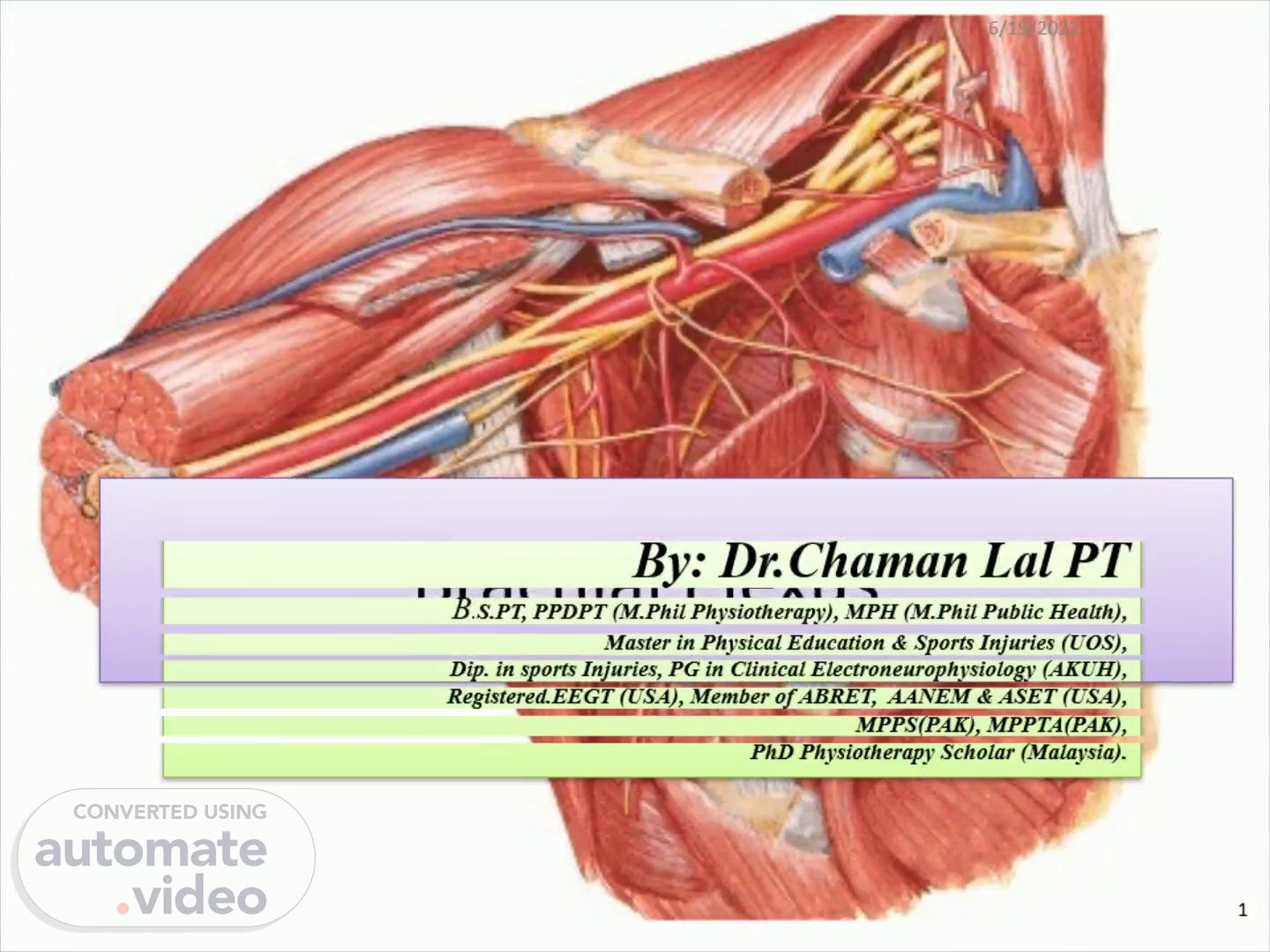
Page 1 (0s)
Brachial Plexus. Master in Physical Education & Sports Injuries (UOS),.
Page 2 (24s)
Brachial plexus: A network of spinal nerves that originates in the back of the neck, extends through the axilla (armpit), and gives rise to nerves to the upper limb..
Page 3 (41s)
1. 3. Brachial Plexus By: CK. 6/19/2022.
Page 4 (48s)
Pcsterior Ot digastnc Sterrxxleid«xnastoid P osterior Triangle of Neck tplly Of TracGus Of omohyoid Maru'ible Anterior belly of digastric Anterior Triangle of Neck.
Page 5 (56s)
BRACHIAL PLEXUS. 5. Brachial Plexus By: CK. 6/19/2022.
Page 6 (1m 6s)
brachialplexus. Brachial Plexus By: CK. 6. 6. 6/19/2022.
Page 7 (1m 14s)
Vertebras & Spinal Nerves. TIE CERVICAL NERVES Head and Neck oelt0idS, Biceps Wrist Extenders Triceps Hana THORACIC NERVES nest Muscles abdominal Muscles LUMBAR NERVES Leg Muscles SACRAL NERVES Bowel. Bladder sexual Function.
Page 8 (1m 29s)
Flexion/Extension of the cervical spine is at; Atlanto-occipital joint Rotation of the cervical spine is at; Atlanto-axial joint.
Page 9 (1m 41s)
vertebrae. atlas1ax. Intervertebral foramen. Atlas (C1).
Page 10 (1m 54s)
Terms To Define?. Horns in Spinal Cords..? Ventral/Anterior Horn Lateral Horn Dorsal/Posterior Horn Root…? Ventral Root Dorsal Root Spinal Nerve…? Ganglion…?.
Page 11 (2m 11s)
“Horns” In The Spinal Cord. In the spinal cord the gray matter is present inside and white matter is towards outside while in brain this distribution is vice versa. On cross section of the spinal cord gray matter in spinal cord presents butterfly like structure form which some projections like “Horns” are seen termed as: Ventral/Anterior Horn→ Lateral Horn → Dorsal/Posterior Horn→.
Page 12 (2m 36s)
Terms To define….cont’d. Root Ventral/Anterior/Motor/Efferent Root: It arises from Ventral or Anterior horn of the Spinal Cord and carries only motor fibers. Dorsal/Posterior/Sensory/Afferent Root: It arises from Dorsal or Posterior horn of the Spinal Cord and carries only sensory fibers. Spinal Nerve: The dorsal and ventral roots, arising from each spinal cord segment, fuse within the intervertebral foramen to form a mixed spinal nerve..
Page 13 (3m 0s)
Dorsal primary ramus (to skin and muscles of back) Ventral primary ramus Dorsal (= posterior) horn of spinal gray matter Lateral horn (preganglionic sympathetic- neurons; only in segments Tl-L2) Ventral (z anterior) horn. (Contains motor neurons) Dorsal psterior) root Ventral Dorsal root ganglion Spinal nerve Gray ramus communicans Sensory fiber Postganglionic sympathetic innervation (glands, blood vessels) Motor axon (to skeletal muscle) White ramus communicans Fig. 51. A thoracic segment of the (= anterior) spinal cord, showing sensory and root motor neurons and the connections of a paravertebral sympathetic ganglion. Sympathetic (paravertebral) ganglion.
Page 14 (3m 18s)
Terms To define….cont’d. Ganglion: Collection of the nerve cell bodies is called “Ganglion” e.g., Dorsal Root Ganglion “DRG”, sympathetic paravertebral ganglion. Ramus (Pl. rami) The spinal nerve when exits from the vertebrae it divides into two branches termed as; Ventral/Anterior Primary Ramus Dorsal/Posterior Primary Ramus Ventral/Anterior Ramus: It is the ventral or anterior division of the spinal nerve out side the vertebrae. It supplies to the skin, muscles on anterior body & viscera etc. Dorsal/Posterior Ramus: It is the dorsal or posterior division of the spinal nerve out side the vertebrae. It supplies to the skin & muscles of the back..
Page 15 (3m 51s)
Spinous process Foramen (filled with adipose tissue) Inferior articular process Superior articular process Posterior tubercle of transverse process Anterior tubercle of transverse process Foramen transversium Vertebral body Meninges Gray matter White matter Dorsal root Ventral root Spinal nerve Nucleus pulposus Disc annulus.
Page 16 (4m 2s)
Dorsal Ventral rcot (spinal) ganglion Spinal nerve Dorsal ramus Of spinal nerve Ventral ramus of spinal nerve nerve) (a) Anterolateral view Communicating rami Ganglion Of sympathetic chain of splanchnic nerve.
Page 17 (4m 11s)
All plexus i.e., Cervical, Brachial & Lumbo-sacral are formed by the fusion of Ventral or Anterior Primary rami of the spinal nerves. Brachial Plexus are formed by anterior primary rami of C 5 , C 6 , C 7 , C 8 , T 1 spinal nerves . Now these are termed as Ventral/ Anterior Spinal Nerve Roots. Brachial Plexus has the following relations to the surrounding structures: The roots lie between the anterior and middle scalene muscles. The trunks traverse the posterior triangle of the neck. The divisions lie behind the clavicle. The cords lie in the axilla, and are named according to their relationship with the Axillary artery..
Page 18 (4m 41s)
6/19/2022. 18. Brachial Plexus By: CK.
Page 19 (4m 50s)
C5 C7 ROOT TRUNK DIVISION CORD D c RANCHES Axillary Nerve Radial Nerve Median Nerve Ulnar Nerve.
Page 20 (4m 58s)
brachial-plexus-vs-axillary. 20. Brachial Plexus By: CK.
Page 21 (5m 6s)
05 08 First rib Trapezius Erb's Palsy Clavicle Axillary nerve (nerve to deltoid) Humerous Nerve to biceps Radial nerve (triceps, wrist and finger opening) Median nerve (hand function) Ulnar nerve (hand function).
Page 22 (5m 15s)
Nerve Roots C5,6,7,8,T1 Trunks Upper Middle Lower Divisions Anterior Posterior Cords Lateral Medial Posterior.
Page 23 (5m 28s)
Brachial Plexus…..cont’d. Roots : These are constituted by the anterior primary rami of spinal nerves C 5 , C 6 , C 7 , C 8 , T 1 . Trunks : Upper Trunk: It is formed by the combination of C 5 & C 6 spinal nerve roots. Middle Trunk: It is formed by the continuation of the C 7 spinal nerve root. Lower Trunk: It is formed by the combination of the C 8 & T 1 spinal nerve roots. The trunks traverse the posterior triangle of the neck..
Page 24 (5m 52s)
`. plexus. 24. Brachial Plexus By: CK. 6/19/2022.
Page 25 (6m 0s)
Brachial Plexus…..cont’d. Divisions: Now each trunk give rise to two divisions i.e., 1. Anterior Division 2. Posterior Division The divisions lie behind the clavicle. Cords: Lateral Cord: It is formed by the combination of Anterior divisions of Upper & Middle Trunks . Medial Cord: It is formed by the continuation of anterior division of Lower Trunk . Posterior Cord: It is formed by the combination of all three posterior divisions of Upper , Middle & Lower Trunks..
Page 26 (6m 26s)
Upper Middle Lower Trunks flexor extensor Anterior Divisions Posterior Plan of the Brachial Plexus Lateral Cord Postenor Cord Medial Cord Cords.
Page 27 (6m 35s)
Brachial Plexus…..cont’d. The cords lie in the axilla, and are named according to their relationship with the Axillary artery. Branches: Branches of the Roots: Long Thoracic Nerve (C 5 , C 6 , C 7 ) Dorsal Scapular Nerve (C 5 ) Branches of the Trunks: Suprascapular Nerve (C 5 , C 6 ) Nerve To Subclavius (C 5 , C 6 ).
Page 28 (6m 53s)
Branches of Brachial Plexus…..cont’d. Branches of the Cords: Lateral Cord (C 5 , C 6 , C 7 ) : It gives Three branches; L ateral pectoral Nerve M usculocutaneous Nerve L ateral root of Median Nerve Medial Cord (C 8 , T 1 ): It gives Five branches; M edial Pectoral Nerve M edial Cutaneous Nerve of Arm M edial Cut.N. of forearm (Medial Antibrachial Nerve) M edial Root of Median Nerve U lnar Nerve.
Page 29 (7m 15s)
Branches of Brachial Plexus…..cont’d. Posterior Cord (C 5 , C 6 , C 7 , C 8 , T 1 ): Upper S ubscapular Nerve Lower S ubscapular Nerve T horacodorsal Nerve (Nerve To latissimus dorsi) A xillary Nerve R adial Nerve Medial Antibrachial→ Medial Cord Lateral Antibrachial→ Continuation of Musculocutaneous Nerve → Lateral Cord.
Page 30 (7m 32s)
Posterior scapular nerv Long thoracic nerve Suprascapular nerve Subclavian nerve Posterior cor Lateral cord Axillary nerve Subscapular nerve Thoracodorsal nerve Radial nerve Musculocutaneous nerve Medial and lateral pectoral nerves Median nerve Ulnar nerve aedial cutaneous antebrachia Medial brachia cutaneous nerve Medial cor Roo ts Trunks Anterior divisions Posterior divisions.
Page 31 (7m 44s)
Main Terminal Branches. 31. Musculocutaneous Nerve.
Page 32 (8m 4s)
c BRANCHES TRUNKS DIVISIONS SUPRASCAPULAR K. UPPER CORDS MtOOLE LONG THORACIC N. MEDIAL PECTORAL N- MEDIAL BRACHIU CUTANEOUS N. MtObU ANTEBRACHIAL CUTANEOUS N..
Page 33 (8m 15s)
33. Brachial Plexus By: CK. 6/19/2022.
Page 34 (8m 22s)
Roots: C7. C8. Trunks: upper. Anterior divisions posterior Cords: posterior. Major branches: Axil lary nerve nerve Med i an nerve nerve Subclavian upper and Suprasca,pu'ar nerve Posterior cord AX nerve Lateral cord Radial nerve cuta Medial and lateral pec t oral Medial antebrachial cuta neous nerve pper Middle trunk Media' cord Medial brachial Anterior view.
Page 35 (8m 37s)
October 8, 2008 Karl Merk, the world's first person to receive a complete double arm transplant. Farmer Merk who received the world's first complete double arm transplant is recovering well and able to perform some basic tasks. It still could take up to two-years until he re-learns how to use his hands. Doctors spent 15 hours on July 25-26 grafting the donor arms onto the body of 54-year-old Karl Merk, who lost his own just below the shoulder in a farm accident involving a combine six years ago..
Page 36 (9m 8s)
First they exposed the muscle, nerves and blood vessels to be connected. Before the bones of the donor could be cut, blood vessels in the teen’s arms were filled with a cooled preservation solution. Both arms were then removed exactly at the point matching Karl’s arm stumps.The bones were joined, then arteries and veins to ensure blood circulation as quickly as possible. The surgeons then attached the muscles and tendons, then the nerves and finally the skin. There had been no sign that Karl’s immune system was rejecting the foreign tissue and his scars were healing well..
Page 37 (9m 41s)
http://www.telegraph.co.uk/news/worldnews/europe/germany/Worlds-first-double-arm-transplant-patient-delighted.html.
Page 38 (10m 8s)
References:. Snell's Text Book of Anatomy “Brachial Plexus” 23 rd Ed. Human Anatomy “Upper Limb& Thorax Anatomy” Vol.I By: B.D Chaurasia. “Anatomy & Physiology” By: Inderbir Singh, JP Brothers Publication. “ESSENCE” (A Review Book of Gross Anatomy) 6 th Ed. Dow Promotion Cell Publication. Electromyography & Neuromuscular Disorders” By David C.Preston & Barbra E.Shapiro 2 nd Ed. www.telegraph.co.uk/news/worldnews/europe/germany/Worlds-first-double-arm-transplant-patient-delighted.html.