MedCom_TraumaticBrainInjury_1E_9789390553983_15,2x22,9_(6x9')SC_3Seiten_k8.indd part 2
Scene 1 (0s)
[Audio] 2 Epidemiology of TBI G. Gururaj Traumatic Brain Injury in India Traumatic injuries have emerged as a major public health problem in India in recent years. The rapid urbanization, unprecedented motorization, growing industrialization, and increasing impact of media along with changing lifestyles and values of people in the absence of safety policies and programs have contributed to this scenario. Even as India has made significant strides in the prevention and control of infectious, nutritional, and communicable diseases the disease patterns and profiles have changed, thus increasing the contribution of injuries to death and disability. Consequently, injuries have become a major cause of mortality, morbidity, and disability in India. Among the various types of injuries, traumatic brain injury (TBI) is at the forefront, resulting in a significant number of deaths, hospitalizations, disabilities, and socio-economic losses. TBIs have a huge impact on individuals and families that represent an increasing burden on society at large. TBIs often affect young and productive members of society, and since most of them survive, they not only become unable to care for their families, but rather become a burden on them. The government is simply unable to deal adequately with this growing epidemic. The importance of the magnitude of the problem due to TBI in India, has always been underestimated due to lack of proper research and good quality data. The development of robust epidemiological data helps in planning preventive strategies. The knowledge of the incidence, severity and the mechanism of injury, allow us to design appropriate health care services from subacute and emergency medicine to neurorehabilitation and also determines the training needs of the healthcare workforce. It also gives us an estimate about future socioeconomic needs to minimize the burden on wider society and governments. Incidence and Mortality The current assessment of the frequency of TBIs in India is limited due to lack of good quality population-based information in this area. There have been no large-scale epidemiological studies in the country. Apart from absence of community-based epidemiological studies, trauma registries as seen in highincome countries (HICs) do not currently exist in India. Further, the available data from the existing national sources of health and police sectors have inherent limitations due to inconsistent reporting. A small number of studies.
Scene 2 (2m 47s)
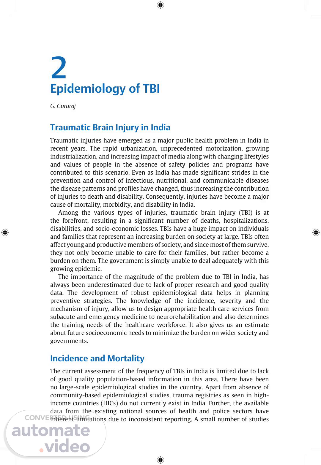
[Audio] National Guidelines for the Management of Traumatic Brain Injury 10 TBIs IN INDIA, 2014 Deaths − 500,000 Hospital ad. − 30,000,000 Minor care − 100,000,000 Fig. 3 Data (2014) from National Mortality Survey for traumatic brain injury (TBI) in India. from individual medical centers provide a limited snapshot of the problem but cannot be accurately generalized in this vast and diverse country. � Estimates from the Global Burden of Disease, World Health Organization, and few independent estimates confirm these observations by reporting more than a million deaths and nearly 50% of them being TBIs.1,2 Further, some of the population-based verbal autopsy studies in India inform that nearly 13 to 18% of deaths in the community are due to injuries. � In a nationally representative mortality survey of 1.1 million homes, it was estimated that nearly 1 million injury deaths occur every year and 60% of them would have an injury to the brain. With this observation it is estimated that there could be nearly 500,000 deaths every year due to TBIs in India (Fig. 3). � Interestingly, the case fatality rates of TBIs, especially in apex institutions and advanced trauma care centers, seem to have reduced due to aggressive management practices. In addition, the availability of specialized manpower and diagnostic-managerial facilities would have contributed to this process. However, this is not true of all other centers in the country. Nonfatal TBIs Several hospital-based studies indicate that nearly half of the injury deaths and hospitalizations have an injury to the brain or spine at admission time, and consequently, it is estimated that about 500,000 TBI-related deaths occur every year in India. Furthermore, for every death nearly 30 to 50 people are hospitalized and consequently it is estimated that a minimum of 15 million moderate-to-severe TBIs are seen in India.3 A recent national review of road safety in India, based on data from the 5-year Bangalore Road Safety and Injury.
Scene 3 (5m 21s)
[Audio] Epidemiology of TBI 11 Prevention Program, has indicated that nearly 20% of ER Registrations, 10% of admissions, and one-third of hospital deaths are due to injuries and more than half of them have a TBI.4 The prevalence of TBIs is not clearly known and a population-based study undertaken in Bangalore reported a prevalence of 94/100,000 population in rural Bangalore. External Causes of TBIs Among the various external causes, road traffic injuries, falls, workplace injuries (industrial/agricultural), violence, sports injuries, and those resulting during disasters account for the majority of TBIs. The problem, profile, and pattern of TBIs due to these external causes vary and are influenced by several macro and micro factors. Epidemiological studies of neurotrauma indicate that road traffic injuries account for 60% of TBIs with falls and assault making up most of the rest. The NIMHANS TBI registry indicated that 60% of TBIs were due to road crashes, 25% due to falls, 10% due to assaults/violence, and the rest due to other miscellaneous causes. With road deaths increasing at an annual rate of 5 to 8% a year, the number of TBIs is also likely to increase at a corresponding pace in the coming years. As per the WHO, road crashes will be the fifth leading cause of death in India by 2030. Data from NCRB also indicate that the economically more progressive Southern states of India have a higher incidence of road crashes as compared to other states. Indian highways that are at a stage of rapid expansion are known to contribute for nearly 40% road deaths and injuries in India with variations seen among different states. Most significantly data indicate that in India pedestrians, two-wheeler riders and pillions, and bicyclists constitute the majority of road traffic victims in India, as compared to high-income countries where motor vehicle occupants make up a much larger proportion. The precise causes for the high incidence of road traffic accidents in India are still to be fully delineated. However, available research indicates that some of the risk factors include road design and operating factors, safety features of the vehicles, nonuse of helmets, seat belts, and child restraints, drinking and driving, driving at excessive speeds, poor visibility, the lack of pedestrian sidewalks, and discipline. Patterns of TBIs Limited data from India indicate that as seen in other parts of world, cerebral concussions, contusions, skull fractures, and traumatic intracranial hemorrhages (epidural, subdural, and intracerebral) are the usual injuries seen.5 The severity distribution indicates that at least 60% of TBIs are classified as mild, 20% are moderate, and 15 to 20% are severe in nature. This proportion of mild TBIs is usually reported as 80% in western studies—perhaps because milder injuries do not always make it to tertiary centers in India..
Scene 4 (8m 47s)
[Audio] National Guidelines for the Management of Traumatic Brain Injury 12 Studies have also indicated that nearly a quarter of the injured persons are likely to have polytrauma and require prompt and intensive management. Disability It is well acknowledged that nearly all patients with severe TBI, 50% with moderate TBI, and 10 to 20% of those with mild TBI need rehabilitation services including physical, psychosocial, vocational, and economic rehabilitation. Based on available limited data, it is estimated that nearly 5 million Indians with a TBI require rehabilitation services.6 Trauma care services in India are evolving in India, even though distinct differences exist in availability, accessibility, and affordability between urban and rural areas. Apart from a major focus on primary prevention of RTIs, trauma care services that integrate emergency, in-hospital, and postdischarge care built on a combination of required human resources, facilities, and services that provide evidence-based care can significantly reduce deaths and disabilities.7 The high burden of injuries and TBIs in India needs the urgent attention of policy makers, professionals, and political leaders, as global evidence has clearly demonstrated that these are eminently predictable and preventable. References 1. Gururaj G. Epidemiology of traumatic brain injuries: Indian scenario. Neurol Res 2002;24(1):24–28 2. World Health Organization. Injury prevention and control. An epidemiological survey of injuries in area of Municipal Corporation of Delhi, New Delhi. SEAinjuries-5, 2003 3. Gururaj G. Road traffic deaths, injuries and disabilities in India: current scenario. Natl Med J India 2008;21(1):14–20 4. Gururaj G. Epidemiology of injuries—a population based survey in Bangalore. In: Proceedings of the 6th World Conference on Injury Prevention and Control. Montreal; 2002 5. Hyder AA, Wunderlich CA, Purvanachandra P, et al. The impact of traumatic brain injuries: a global perspective. Am J 2007;22(5):341–353 6. Gururaj G. An epidemiological approach to prevention: pre-hospital care and rehabilitation in neurotrauma. Neurol India 1995;43(3):95–105 7. Gururaj G, Gautham MS. Advancing road safety in India: implementation is the key. Bengaluru: National Institute of Mental Health & Neuro Sciences; 2017. Publication Number 136. https://nimhans.ac.in/wp-content/uploads/2019/02/UL_BR_m010-11_ Main-rprt_FINAL.pdf. Accessed on April 10, 2021.
Scene 5 (12m 24s)
[Audio] 3 Prehospital Care Guidelines V. D. Sinha and Amit Chakrabarty Brain resuscitation should be started at the earliest as the traumatized brain is very susceptible to hypoxia and ischemia, and this makes prehospital care a crucial link in the chain of trauma care. Prehospital and emergency care has been a major issue in India, and it has been estimated that up to 20% of injured patients die after accidents because of inadequate treatment prior to hospitalization. The first 60 minutes after the trauma have been identified as the critical period for transporting patients from the scene of accident to a health-care center, preferably a trauma center and has been called as the "golden hour". The benefits of providing definitive resuscitative care within this early window have been widely acclaimed worldwide. Several studies in trauma literature have found reduced odds of dying with reduced prehospital times and first-aid care. The 'Golden hour concept' is now being fortunately getting accepted, at least in some metropolitan cities in India. The morbidity and mortality of severe head injury could be remarkably reduced by simply securing and maintaining the airway and stopping any external bleeding before arrival at the hospital. In the Indian setting, the prehospital guidelines are meant for any personnel involved in the prehospital response team such as the retrieval individuals, transporters, emergency room staff, medical personnel, specialists and health care workers. The training of the prehospital emergency personnel is often limited, and this can make a tremendous difference, especially when transfer times are long. There is a significant need to increase basic life support (BLS) emergency medical service (EMS) on site, and improved use of advanced life support (ALS) EMS systems. Ideal Circumstances In an ideal situation three critical tasks must be rapidly performed by prehospital providers caring for trauma victims: � Examination with recognition of severe injuries and injuries with potential to cause rapid decompensation. � Stabilization and transport to a hospital capable of addressing the identified injuries. � Performance of an efficient triage with multiple victims. Triage of trauma victims is the process of rapidly and accurately evaluating patients to determine the extent of their injuries and the appropriate level of medical care required. The goal is to transport all seriously injured patients to medical facilities capable of providing appropriate care, while avoiding.
Scene 6 (15m 13s)
[Audio] National Guidelines for the Management of Traumatic Brain Injury 14 unnecessary transport of patients without critical injuries to trauma centers. Performing full spinal immobilization is prudent; prolonging scene time to initiate intravenous lines, bandage nonhemorrhaging wounds, or splint minor fractures is unnecessary and potentially deleterious. Airway and Breathing The airway must be assessed first in all trauma patients: � If the patient can talk then the airway does not need immediate intervention. � Noisy breathing is a sign of airway compromise requiring immediate intervention: Use a jaw thrust, remove foreign bodies from the mouth, perform an oral suction, and then insert an oral airway. It is important in the prehospital setting to protect the airway of head-injured patients in the least invasive manner possible. An oral or nasal airway (with oral suction) is often sufficient if the airway is compromised, and the patient is breathing adequately. If the breathing is inadequate, then a definitive airway is indicated. If transport time is brief, often bag-mask ventilation (BMV) alone is sufficient, but in most cases with inadequate breathing either a laryngeal mask airway (LMA) or endotracheal intubation will be required. At all times care must be taken to maintain cervical immobilization as best as possible. High-flow oxygen must be supplied to all trauma patients as soon as the airway and breathing are secured. Oxygen saturation should be continuously monitored with a pulse oximeter and the saturation maintained >90%. Blood Pressure All efforts must be made to keep the systolic blood pressure >90 mmHg in adult trauma patients. � Control external hemorrhage with compression and bandaging. � Splint long bone fractures, especially in the lower limbs. � The decision to start IV fluids on the scene depends on the severity of hypotension and the estimated time to hospital. Fluids should be isotonic and not contain dextrose and administered at the maximum possible rate. If there is no hypotension, patient transfer should not be delayed by starting IV lines. The blood pressure should be monitored frequently during resuscitation and transport. Cardiac Arrest If there is no palpable pulse, then basic life support must be started and continued without interruptions until the patient is taken over by the hospital team..
Scene 7 (17m 57s)
[Audio] Prehospital Care Guidelines 15 Glasgow Coma Scale Score Prehospital measurement of the Glasgow Coma Scale (GCS) is a reliable indicator of the severity of TBI but should not be assessed until oxygenation and blood pressure have been normalized. Repeated assessment should be performed to identify improvement or deterioration over time. The adult protocol for standard GCS measurement can be followed for children over 2 years of age. In preverbal children, the pediatric GCS should be employed, with a full verbal score of 5 assigned to infants cooing or babbling. Pupil Examination Pupils should be assessed in the field for use in diagnosis, treatment, and prognosis. Pupillary examination should be done only after primary resuscitation, and the size and reaction of the pupils must be assessed. Left and right pupillary findings must be recorded separately, and asymmetry is defined as >0.4 mm difference in diameter. Limited Resources In many locations in India, the personnel available for retrieval of trauma patients do not have the training or the equipment to provide the interventions described above. It is important that these personnel are made aware of what they can do, besides just transporting the patient. These basic interventions for airway and blood pressure can be performed with no/minimal equipment and will result in significant improvements in the outcome of the patient. Protocols It is important that each area formulate realistic protocols for the situation on the ground in that region. For example, having a protocol that requires intubation as the intervention for a compromised airway when the majority of personnel do not have the necessary training will only result in no intervention being carried out and the airway remaining compromised during the entire transport. If they are taught that the minimal intervention is still better than none it is more likely that the patient will benefit. This is often not done because the organization will feel that accepting these lesser interventions is compromising their standards, but this will actually improve patient outcomes and must be seen as necessary. Protocols are necessary regarding patient transport to appropriate facilities depending on the patient's clinical status; this is a regional decision and cannot be prescribed from a central organization..
Scene 8 (20m 23s)
[Audio] National Guidelines for the Management of Traumatic Brain Injury 16 Airway and Breathing The process of assessment does not change—if the patient is not talking then the airway is at risk. � If there is no noisy breathing and the chest is moving adequately no intervention is required, but this should be regularly checked during transport. � If the breathing is noisy, clear out the mouth with a jaw thrust, perform an oral suction, and insert an oral airway. Repeat suction as often as necessary. � If suction or artificial airway are not available and the breathing remains noisy, turn the patient semiprone—this will move the tongue away from the back of the throat and allow secretions or blood to flow out of the mouth. � If the breathing is inadequate after clearing the airway and a definitive airway is not possible, attempt bag-mask ventilation and transport the patient as rapidly as possible to the nearest hospital where a definitive airway can be placed. In all cases high-flow oxygen must be started soon after these interventions. Blood Pressure If blood pressure measurement is not possible, the retrieval personnel can be taught to assess pulse rate and volume. � Control all external hemorrhage with compression and bandaging. � If the expertise to start IV lines is not available and the blood pressure is low the patient must be transported to the nearest medical facility for this purpose before further transport to a trauma center..
Scene 9 (22m 9s)
[Audio] 4 Hospital Care Guidelines Sumit Sinha and Mathew Joseph Levels of Care Traumatic brain injury (TBI) patients are treated in both government facilities and private hospitals. Government hospitals are generally overburdened, with inadequate manpower, resources, and equipment for the patient load. Available personnel and their skills often do not match the needs of the patients, and the concept of a dedicated trauma team has generally not been established. Small hospitals mushrooming across India label themselves as trauma centers but provide inadequate care with high mortality rates. Privately provided high-level care is expensive and therefore unaffordable to a majority of trauma patients. Hospitals need to be categorized by the resources they have, to ensure that patients are appropriately managed at the level of care they need. Patients with significant injuries should not be retained at smaller hospitals, and at the same time there should not be unnecessary referrals and overcrowding of the higherlevel hospitals. � Level 1 hospitals: should have round-the-clock computed tomography (CT) scan facilities, neurosurgical and neurocritical care, and the ability to manage major trauma of other systems. They should be able to handle head injuries of any severity. � Level 2 hospitals: should have round-the-clock CT scan facilities and a neurosurgeon on call available within an hour. These hospitals usually have limited ICU facilities and full-fledged neurocritical care is not possible. These hospitals should treat moderate head injuries, as well as minor head injuries requiring neurosurgery. � Level 3 hospitals: are hospitals where round-the-clock CT scan and most other trauma services are available, but no neurosurgery is available. There should be an established method of obtaining a remote neurosurgical consultation. Patients with moderate and mild head injuries can be managed here depending on the CT findings after consultation with the remote neurosurgeon. � – Any abnormal CT scan should be referred to a higher level if transfer time to neurosurgical care is more than 2 hours. � – Liability issues for the consulting neurosurgeon need to be sorted out. � Level 4 hospitals: do not have CT scan facilities but are able to stabilize airway, breathing, and circulation (ABC) and might have other trauma services. Patients with TBI who do not require CT scans can be treated at these hospitals..
Scene 10 (24m 49s)
[Audio] National Guidelines for the Management of Traumatic Brain Injury 18 Emergency Management At arrival in the hospital the immediate focus must be on the ABC, and any assessment of the TBI must begin only after the necessary interventions for the ABC have been initiated. Monitoring Airway Judging the patency of airway in a nonintubated patient is always clinical. � Causes of airway compromise in HI: � – Altered consciousness—tongue fall back. � – Vomitus. � – Blood, facial injuries. � Signs of airway compromise: � – Does not talk. � – Noisy breathing. � – Obvious facial injury. � Indicators of potential airway compromise: � – Use of accessory muscles. � – Neck injury. � – Short neck. Airway assessment in a patient with a secured artificial airway. � Position: � – Auscultation. � – Chest radiograph. � Patency: � – Effort of breathing. � – Ease of passing suction catheter. � – Airway pressures if on ventilator. In all head injury patients high-flow oxygen should be administered immediately after securing the airway. Breathing The adequacy of spontaneous breathing is again primarily clinical, with laboratory assistance. � Causes of inadequate breathing: � – Obstructed airway. � – Impaired effort: � Severe neurological injury. � Drug induced (sedation, muscle relaxants)..
Scene 11 (26m 25s)
[Audio] Hospital Care Guidelines 19 � – Chest pathology: � Aspiration. � Chest trauma. � Clinical signs of inadequate ventilation: � – Rapid or very slow respiratory rate. � – Use of accessory muscles. � – Poor chest expansion / air entry. � Accessory means of assessing breathing: � – Arterial blood gas. � – Low SpO2 on a pulse oximeter (normal SpO2 while on supplementary oxygen does NOT indicate adequate breathing). Breathing in a patient on a ventilator is assessed clinically and with assistive technology. � Clinical: � – Chest movement. � – Auscultation. � ICU technology: � – Ventilator parameters. � – Pulse oximetry. � – End-tidal CO2. � – Arterial blood gas. Blood Pressure Assessment of adequacy of circulation will depend on where the assessment is being done: � Causes of hypotension: � – Blood loss—external or internal. � – Chest trauma: � Tension pneumothorax. � Cardiac injury or tamponade. � – Cervical spine injury (hypotension without tachycardia). � – Patient almost brain dead. Emergency room signs of impaired circulation: � Pulse—tachycardia, poor volume. � Blood pressure. � Capillary refill >2 seconds, cold extremities. � Other evaluation: � – Auscultation for pneumothorax or tamponade. Monitored area (in ER or high care area): � Heart rate..
Scene 12 (28m 10s)
[Audio] National Guidelines for the Management of Traumatic Brain Injury 20 � Noninvasive blood pressure. � Invasive blood pressure—if significant instability present or on vasoactive drugs. Intervention Airway Assessment and intervention should ideally be done as follows: � Talk to the patient—if he answers airway is secure. � If he does not speak perform a jaw thrust, remove foreign bodies from the mouth, perform an oral suction, and then insert an oral airway. � If even after this breathing is noisy, or there is significant blood in the oral cavity the patient needs a definitive airway. � – Endotracheal intubation/laryngeal mask airway is the primary option. � – If not possible a cricothyroidotomy should be done (a tracheostomy is not an emergency surgical intervention). � Connect high-flow oxygen after securing airway. � If a definitive airway was not initially necessary, frequent reassessment of the airway is mandatory. Breathing If breathing is inadequate the patient will need a definitive airway (unless the reason is rapidly correctable such as a tension pneumothorax). Inserting an endotracheal tube will usually require sedation and often muscle relaxants, following which breathing has to be assisted. This can be done manually or (where available) with a ventilator. Care must be taken to: � Supply supplementary oxygen. � Avoid hyperventilation (common with manual ventilation). Blood Pressure Hypotension is extremely damaging in head injury, and prevention/rapid correction is very important. � If blood pressure is low or patient has tachycardia with borderline blood pressure, start large caliber peripheral lines in both antecubital fossae and administer dextrose-free crystalloids at maximum rate. � Send a cross match for emergency blood transfusions. � Control hemorrhage: � – External. � – Abdomen—FAST and urgent surgical consultation if positive. � – Pelvis—apply binder. � – Femur—splint..
Scene 13 (30m 23s)
[Audio] Hospital Care Guidelines 21 � Continue to administer crystalloids rapidly and monitor pulse and blood pressure. � After more than one liter of fluid is administered ringer lactate is preferred. � If the blood pressure does not respond to crystalloids alone and crossmatched blood is not ready O-negative blood may be transfused. � Surgical control of hemorrhage may be needed if the circulation can still not be stabilized. � Once hemorrhage is controlled, vasoactive drugs may be used to raise the blood pressure if suitable monitoring is available. The treating personnel should examine and make every effort to exclude, especially in an unconscious patient, the easily overlooked causes of hypotension such as cervical spine injury, abdominal injury, pelvic fractures or fractures of long bones. Limited Facilities If the sequence of interventions described above cannot be performed due to lack of facilities, then the following steps should be taken before referring the patient to a higher center: Assessment of ABC remains unchanged and should be documented for the benefit of subsequent management. Airway If a definitive airway (endotracheal intubation/LMA/surgical airway) is not possible then: � Insert an oral airway and perform suction. � If frequent suction is needed or breathing is still noisy after inserting the airway, turn the patient semiprone so that oral secretions/blood flow out of the mouth. � If an oral airway is not available, turn the patient semiprone. Breathing � If a definitive airway is possible and breathing inadequate: � – Connect to ventilator. � – If ventilator not available refer to a higher center. � – If this is not possible continue manual assistance and correct cause of impaired breathing until breathing stabilizes. � If a definitive airway is not possible then secure airway as best possible, treat any easily treatable cause of impaired breathing and refer urgently. Circulation � If blood pressure is low, start rapid infusion of crystalloids..
Scene 14 (32m 32s)
[Audio] National Guidelines for the Management of Traumatic Brain Injury 22 � If facilities for transfusion/treatment of extracranial injuries are not available: � – Control hemorrhage as well as possible. � – Refer urgently to a higher center while continuing rapid infusion of crystalloids en-route. Management of TBI The principal focus of treatment in TBI is to limit secondary brain injury. This treatment is therefore aimed at optimizing oxygenation, blood pressure, intracranial pressure, and maintenance of adequate cerebral perfusion. It is also important to manage temperature, glucose, seizures, and other causes of potential secondary brain insults. All personnel involved in monitoring a head injury patient must be trained to assess the Glasgow Coma Scale and pupils. The following sections deal with the treatment of TBI in a hospital that has the necessary personnel and equipment for the purpose. Criteria for Admission The decision on where to admit the patient (emergency ward, regular ward, or ICU) will depend on the availability of facilities for monitoring and treatment at each institution, and these policies should be clearly defined to avoid any confusion. The following are the criteria for admitting a patient with TBI: � History of loss of consciousness or amnesia. � Age >65. � Glasgow Coma Scale (GCS) score <15. � Deteriorating GCS score. � Focal neurological deficits. � Posttraumatic seizure. � Suspected skull fracture or penetrating injury. � Drug or alcohol intoxication. � Patient on antiplatelet drugs or anticoagulants. � History of coagulopathy. � Other significant injuries. � Cerebrospinal fluid leak. � No responsible caregiver after discharge. � Persistent headache, photophobia, nausea, or vomiting. Criteria for Performing a CT Scan A CT scan should be performed when breathing and hemodynamic status allows transport. � GCS score less than 13 on initial assessment..
Scene 15 (34m 4s)
[Audio] Hospital Care Guidelines 23 � GCS score less than 15 at 2 hours after the injury. � Amnesia for the event. � Suspected penetrating or depressed fracture. � Suspected base of skull fracture (hemotympanum, "panda" eyes, cerebrospinal fluid leakage from the ear or nose, Battle sign). � Posttraumatic seizure. � Focal neurological deficit. � Two or more episodes of vomiting. � Patient on antiplatelet drugs or anticoagulants. � History of coagulopathy. � Persistent headache, nausea, giddiness, restlessness. � Before clearance for anesthesia for other injuries (do not delay emergency surgery to control hemorrhagic shock). � Tense anterior fontanelle (in infants). Indications for a Repeat CT Scan � Deterioration of GCS score. � New onset or worsening focal neurological deficit. � Persistent headache, vomiting, or restlessness. � Bradycardia, hypertension. � Abnormal initial CT scan (repeat at 24 hours or earlier if indicated). Note: There is evidence that the radiation exposure from a CT scan can damage the brain of children <5 years old. Modern CT scanners have pediatric settings to decrease this exposure—make sure they are used. The NTSI does not at this time recommend MRI scans for children with acute TBI. Monitoring Parameters to be monitored outside the ICU: � Pulse. � Blood pressure. � Clinical assessment of airway and breathing. � Temperature. � GCS score. � Pupils. � Other significant injuries/problems of that patient. Frequency of systemic monitoring: � If no instability at admission, then once an hour for 6 hours. � If stable then continue once every 2 hours..
Scene 16 (35m 25s)
[Audio] National Guidelines for the Management of Traumatic Brain Injury 24 Frequency of neurological monitoring (ABC stabilized): � Every half an hour till some sign of neurological improvement. � Then every hour till 12 hours. � Subsequently reduce frequency to every 2 hours if there has been some improvement since admission. Response to deterioration: � Report immediately. � Rapid decision to be made on the need for: � – Repeat investigation. � – Referral. � – Transfer to ICU/OR. ICU monitoring: � Monitoring depends on severity of injury and capabilities of the system. For a patient who is being ventilated, EKG, invasive blood pressure (or frequent automatic noninvasive), pulse oximetry, and end-tidal carbon dioxide measurements should be monitored. � If intracranial pressure (ICP) monitoring is possible then it should be done for: � – All salvageable patients with GCS of 3 to 8 and abnormal CT. � – In patients with GCS of 3 to 8 with normal CT if two or more of the following are present: � Age > 40 years. � Unilateral or bilateral motor posturing. � SBP < 90 mmHg. The gold standard monitoring technique is an intraventricular catheter connected to an external strain gauge. Current intraparenchymal monitors are also accurate, but other techniques such as subarachnoid bolts are no longer accurate enough to recommend. There is intense research into noninvasive monitoring, but it is not yet in general use. Fluid–Electrolyte Management � Patient must be well hydrated, initially with isotonic fluids that do not contain dextrose. � Hydration monitored by: � – Pulse, BP. � – Urine output and color. � – Intake/output chart if possible. � Hyperosmolar agents should be administered via a central lined. � Adjustments must be made for increased temperature/motor activity. � Enteral feeds must be established as early as possible..
Scene 17 (36m 44s)
[Audio] Hospital Care Guidelines 25 Monitoring electrolytes: � Daily measurement of Na/K is ideal. � Mandatory if hyperosmolar therapy is being administered. Anticonvulsants in TBI A loading dose of anticonvulsant should be administered to all patients who require admission. The commonly used drugs are phenytoin, fosphenytoin, and levetiracetam. There is no evidence that administration of anticonvulsants for more than a week is beneficial, and the medication generally should not be continued after discharge unless the patient had seizures during the admission. Seizures Prophylaxis: In patients with seizures, phenytoin is recommended in order to decrease the incidence of early posttraumatic seizures (PTS) generally within 7 days of injury, when the overall benefit is thought to outweigh the complications associated with such treatment.1 Ventilation in TBI Ventilation in TBI should be adjusted to maintain a pO2 of at least 100 mmHg and a pCO2 of around 35 mmHg. The pCO2 should not be lowered below 30 mmHg except in very specific circumstances. Treatment of Raised ICP The basic treatment of potentially raised ICP is very important in all patients and is unfortunately often neglected. � Head must be elevated above heart level. � Neck must be in neutral position. � Cervical collar must be removed as soon as the spine has been cleared. � Airway and breathing must be normal. � Blood pressure must be adequate. � Temperature must be controlled. � Seizures must be prevented. � Sodium must be maintained. When ICP is not monitored: � Therapy for raised ICP should not automatically be started for all head injury patients. There must be some radiological or clinical evidence of possible raised ICP. � Treatment should include measures to lower ICP and also to maintain an adequate blood pressure and thereby an adequate cerebral perfusion pressure (CPP). � Osmotherapy: hypertonic saline, mannitol, or both in combination may be used. Mannitol is more effective in intermittent doses (1–2 g/kg/d) not more than 6 hours apart. Hypertonic saline may be administered either.
Scene 18 (39m 26s)
[Audio] National Guidelines for the Management of Traumatic Brain Injury 26 continuously or as bolus doses to raise serum sodium up to 150 mEq/L (up to 160 mEq/L in children). When ICP is monitored: � ICP elevation above 22 mmHg requires intervention. � CPP must be maintained above 60 mmHg, using vasoactive drugs if necessary. � Response to elevated ICP: � – Confirm accuracy of reading. � – Ensure all systemic factors listed at the beginning of this section are controlled. � – Vent CSF if an intraventricular catheter is in place. � – Increase sedation and analgesia. � – Osmotherapy. � – Administer muscle relaxants. � – Hyperventilate to a pCO2 of 30 mmHg while preparing definitive intervention. � – At some stage, especially before hyperventilation a CT scan must be repeated. � – If ICP is not controlled with all medical measures and there is no lesion that can be surgically removed, then the options for treatment are: � Barbiturate coma (possible only in high-level ICUs). � Decompressive craniectomy. � – Steroids should NOT be used for treatment of raised ICP. Analgesia Adequate analgesia is mandatory in all trauma patients and should be titrated to the severity of the soft tissue and skeletal injuries. Sedation Sedation should be used by experienced personnel. Short-acting medications are preferred. � Emergency room: � – Agitation. � – For procedure. � – Securing airway. � – Splinting, lines. � ICU: � – Ventilation (as an infusion). � – Procedures..
Scene 19 (41m 22s)
[Audio] Hospital Care Guidelines 27 � Non-ICU areas: � – Extreme caution because of decreased intensity of monitoring. � – Drugs—quetiapine/risperidone/haloperidol/midazolam. Infection Prophylaxis There is no indication for administering prophylactic antibiotics in TBI. Antibiotics should not be administered for CSF rhinorrhea/otorrhea, insertion of ICP monitoring device, or for ventilation unless there are other indications of an active infection. Antibiotics can be given for scalp lacerations and compound depressed fractures that do not require surgery. Operative Therapy (Timing, Indications) Decisions regarding the need, timing, and type of surgery are left to the individual neurosurgeon managing the patient. Uncontrolled rise in ICP or clinical signs of herniation are indications for immediate surgery. Discharge and Follow-Up It is important that all patients after a TBI are sent home with a responsible caregiver who understands the symptoms for which the patient may need to return to the hospital. From the emergency room: � If there is no indication for admission or CT scan by the criteria mentioned above the patient can be sent home after any necessary first aid and treatment of other injuries. � If the CT brain is normal, the patient has a normal sensorium and there are no other indications for admission. Discharge after observation/treatment: � This decision has to be made by the treating team. In an ideal situation all patients should return to independent function before being sent home either from the acute care ward or from a rehabilitation facility. In actual practice this is often not possible due to the pressure to admit new cases of acute TBI, and therefore patients are often discharged early. In this situation an assessment must be made on whether the patient requires in-patient treatment at a lower-level hospital or if the family can be trained to take care of the patient at home. Level 1 and level 2 hospitals should reach out to lower-level hospitals to establish systems by which patients can continue to receive medical (nonneurosurgical) care in a hub and spoke model. Discharge advice: � In all cases it is important to give verbal and printed discharge advice to patients with any degree of head injury and their families and caretakers. This advice should include:.
Scene 20 (44m 0s)
[Audio] National Guidelines for the Management of Traumatic Brain Injury 28 � Details of the nature and severity of the injury. � Details about the recovery process including the possibilities of later difficulties or complications. Symptoms that require a return for neurosurgical investigation should be emphasized. � Information on when to return for follow-up and further treatment. � Information about return to everyday activities including school, work, sports, and driving. Reference 1. Carney N, Totten AM, O'Reilly C, et al. Guidelines for the management of severe traumatic brain injury, Fourth Edition. Neurosurgery 2017;80(1):6–15.
Scene 21 (44m 46s)
[Audio] 5 Neurorehabilitation after Traumatic Brain Injury Dhaval Shukla Rehabilitation is the combined and coordinated efforts of a physician supervised multidisciplinary team in helping a diseased person to reach maximum physical, psychological, social, vocational, and educational potential, consistent with his or her physiological or anatomical impairment, environmental limitations, desires, and life plans. About 10 to 15% of patients following mild TBI, 40 to 50% after moderate, and almost 100% after severe TBI have one or the other disability, requiring rehabilitation. Evaluation � A person with TBI should be evaluated and treated for impairments in cognition, vision, speech and language, behavior, swallowing, sensorymotor system, and bowel and bladder function. � Any patient with TBI who has persistent and stable neurological deficit, and who requires medical monitoring and has impairment in two or more key domains should be transferred to an in-patient neurorehabilitation facility. Recommendations for Neurorehabilitation Facilities � There should be provision for inpatient rehabilitation beds, manpower, equipment, and space to provide optimum care in proportion to acute care. � Each Level I center managing traumatic brain injury must have integrated multidisciplinary in-patient rehabilitation services. � It is recommended for Level II center managing TBI to have integrated multidisciplinary inpatient rehabilitation services. � If in-house rehabilitation facilities are not available then a referral to a rehabilitation physician (physiatrist) should be done, and treatment plan should be obtained for rehabilitation in nursing home or at home. Rehabilitation Team 1. Rehabilitation physician (Physiatrist). 2. Neuropsychologist. 3. Speech and language pathologist..
Scene 22 (46m 58s)
[Audio] National Guidelines for the Management of Traumatic Brain Injury 30 4. Physical therapist. 5. Occupational therapist. 6. Rehabilitation nurse. 7. Orthotist. 8. Social worker. 9. Access to other medical specialties as best available in the setting. Recommendations for Nursing Homes Where Rehabilitation Facilities Are Not Available In-Patient Management Disorders of Consciousness � Prognosis is not universally poor in patients with disorder of consciousness. � No patient should be labeled as "vegetative state (VS)" during acute care. � Periodic assessment using Coma Recovery Scale-Revised should be done to know whether the patient is in unresponsive wakefulness syndrome (UWS), minimally consciousness state (MCS), or emerging from MCS. � Prescribe amantadine (100–200 mg bid) for adults with traumatic VS/ UWS or MCS (4–16 wk postinjury) to hasten functional recovery and reduce disability early in recovery. � Predicting a timeline for coma recovery is difficult, as it mainly depends on the severity of the person's brain injury. � The general time periods of recovery are following; however, these may vary from individual to individual: � – Patients who reach an MCS within 3 months have a high likelihood of regaining full consciousness. � – Recovery after TBI is fastest between 3 and 6 months after injury. � – The speech and mobility improve between 6 months and 2 years. � – Recovery slows down or plateaus 2 years after injury. � Patient should be re-evaluated with imaging when the recovery halts or there is regression of recovery process. � Decision to discontinue care should not be made for at least 3 months after injury. � Decision to discontinue care should be made after repeated consultation with neurosurgeon, social worker, psychologist, and family. Skin Care to Treat or Help Prevent Pressure Sores � Evaluate the person's skin daily for pressure sores or evidence of breakdown. � Wash all open sores daily with boiled water and cover wounds with sterile gauze or clean cloths. Topical agents may be used if wounds are.
Scene 23 (49m 32s)
[Audio] Neurorehabilitation after Traumatic Brain Injury 31 infected or there is pus draining or the area is warm to touch with signs of inflammation. � It is important to clean wet or soiled skin immediately after urination or bowel movements. Prolonged contact with urine or feces can cause skin breakdown. � Consider using a pad or pillow stuffed with cotton. The cotton can be thrown away if soiled and the pad or pillow can be reused. � It is important to turn the person in bed every 2 to 3 hours (during day and night), always checking the skin for red marks that occur with prolonged focal pressure. Care to Prevent Choking and Facilitate Breathing � Persons who cannot move spontaneously should lie in a position that helps easy breathing. � Move pillows and blankets away from face (potential suffocation hazard). � Use rolled blankets to raise the head and chest if lying flat. � Use the sitting position for feeding/drinking, and keep the patient upright for an hour after feeding. � If the patient vomits, quickly turn on side to avoid aspiration/choking. Management of Urine and Feces � Make certain that the patient is having regular urination and bowel movements. � Fruits and grains are most helpful for stimulating bowel motility. � Encourage water intake several times a day. � Isabgol is a good stimulant for having regular bowel movements. � Use Vaseline in anus to assist in removing impacted feces. � A temporary catheter may be necessary for urinary retention. � If used, keep the catheter clean. � Urinary catheter tube should not be in between legs. � Regularly check urine for color and smell. � Dark brown urine is a sign of dehydration, and foul-smelling urine may need further evaluation for possible infection. Reducing the Risk of Limb Deformity � Change patient's position regularly; avoid same position for long duration. � Regularly move both lower and upper extremities. � Mobilize joints to full range passively (or actively if possible). � If splints are used to prevent deformities ensure that there is a soft padding to avoid injuries due to splint itself..
Scene 24 (51m 46s)
[Audio] National Guidelines for the Management of Traumatic Brain Injury 32 � Keep a soft padding or a separator in between the limbs, both upper and lower, fingers, to avoid frictional injuries. Spasticity � Spasticity is a common symptom, which requires intervention, especially when it interferes with functional abilities such as mobility, positioning, or hygiene, or when it is the cause of deformity or pain. � Baclofen appears to improve lower extremity spasticity, and its starting dose is 5 mg tid to maximum dose 80 mg/d. � Tizanidine is effective for improving upper and lower extremity spasticity, and its starting dose 4 mg to maximum dose 36 mg/d. Eating and Drinking If the patient has difficulty swallowing: � Make sure the patient is sitting upright. Support head if necessary. � Encourage to take one sip/bite at a time. � Help the patient to lower chin down toward chest during swallowing. � Try giving liquids by spoon or straw. � Ask the patient to swallow two times for each sip/bite. � Ask the patient to take a breath and hold it, then swallow and breathe. � Check between swallows and after the meal to make sure there is no food left in the mouth. � Offer one item of food at a time. � Do not talk while eating or drinking. � Start with baby food, e.g., halwa, khichdi, banana, ganjee, etc. � A caregiver should remain with patient to assure that patient eats and drinks safely. � Assist patient to sit upright for 30 to 60 minutes after eating. � Patients with cognitive difficulty in feeding may require instruction as given to small children with modification of utensils. Sleep and Rest � Ensure that patient gets a quiet but monitored environment to ensure good sleep. Washing and Bathing � Daily washing is essential for hygiene. � Surgical site needs to be washed daily to prevent infections..
Scene 25 (53m 53s)
[Audio] Neurorehabilitation after Traumatic Brain Injury 33 � Patient with poor balance should sit on a chair with a cut-out opening in the seat, and a back strap to bathe. � Water should be just warm as patients may not distinguish between hot and cold water. Walking � Initial walking can be very tiring. � Walk short distances at first and allow the person to rest often. � Allow to stand and get balance before taking any step forward. � Specific sit-to-stand training results in improved abilities. � Conventional gait training with walker or handheld assistance under supervision, with verbal and tactile cues should be done. � Aerobic exercise helps to reduce fatigue and improves social integration, physical independence, levels of spasticity, and overall mental health. Agitation and Destructive Behavior After brain injury, some persons easily become angry, restless, or excited, or behave in a manner that is not socially acceptable. A person may swear or shout very vigorously and be unable to control this very unpleasant behavior. The person may hit, pinch, bite, or break things for no clear reason. � It may be possible to calm the person simply by talking quietly and in a comforting and reassuring manner. � Soft music can help an agitated person to relax. � Move the patient to a quiet room and remain in that environment, away from other people, until they become calm. � Avoid benzodiazepines (diazepam, lorazepam, etc.). � Oral risperidone 1-2 mg/d or quetiapine (after ECG to rule out QTc prolongation) beginning at 25 mg twice a day may be given. Out Patient Management The following issues can be addressed at outpatient level: � Speech and language. � Headache. � Vertigo. � Cognitive rehabilitation. � Psychiatric symptoms. � Continuation of rehabilitation for motor impairments in ambulatory patient..
Scene 26 (56m 4s)
[Audio] National Guidelines for the Management of Traumatic Brain Injury 34 Indications for Referral to a Specialist Neurosurgeon: � Craniotomy site persistent pus discharge (maybe bone flap osteomyelitis). � Cranioplasty for patients who have undergone decompressive craniectomy. � – Usually 3 to 6 months after injury. � – May consider early if patient has recovered and ready to go for work, scalp is sunken, and local headache. � Posttraumatic hydrocephalus: � – Suspect when recovery halts or when patient starts deteriorating after initial recovery. � Chronic subdural hematoma (CSDH): � – Suspect when new-onset headache days after injury. � – New-onset neurological deficits days after injury. � – Suspect when patient starts deteriorating after initial recovery. Neurologist: � Seizure while on antiepileptic drug (consider cognitive side effects of AEDs before adding new drug). � Posttraumatic headache not responding to NSAIDs or amitriptyline. Rehabilitation specialist: � Spasticity. � For pressure sores not responding to dressings and position change. Orthopedic surgeon: � Heterotopic ossification: � – Suspect when painful restriction of joints with swelling. Chest physician: � Pneumonia. � Venous thromboembolism. Urologist: � Recurrent urinary tract infection. � Complication of neurogenic bladder (urinary stone, pyelonephritis). Gastroenterologist: � For percutaneous endoscopic gastrostomy (PEG) if patient cannot be fed orally for many days or in UWS/MCS. Psychiatrist: � Depression. � Psychosis. � Substance abuse prior to or after injury..
Scene 27 (58m 12s)
[Audio] Neurorehabilitation after Traumatic Brain Injury 35 Outcome Assessment � Glasgow Coma Scale during acute care, and at time of discharge. � Disability Rating Scale (DRS) at time of discharge from acute care. � DRS at time of admission and discharge from in-patient rehabilitation. � Glasgow Outcome Scale Extended (GOSE) at 6 months. An app for GOSE is available on Google Play..
Scene 29 (58m 55s)
[Audio] 6 SummaryandConclusionsSumitSinhaIN-HOSPITALCAREINTRAUMATICBRAININJURY(TBI) LevelISevereTBIRound-the-clockcomputedtomography(CT)scan,roundtheclocklaboratoryforbiochemicalevaluation,mobilechestX-rayunit,ECG,mobileCT(ifpossible),physiotherapyservicesinICU,otherspecialistsavailable—pulmonologists,cardioloigsts,nephrologists,endocrinologistsetc.,neurosurgicalandneurocriticalcare,andtheabilitytomanagemajortraumaofothersystemsShouldbeabletohandleheadinjuriesofanyseverityLevelIIModerateheadinjuries,aswellasminorheadinjuriesrequiringneurosergeryRound-the-clockCTscanfacilitiesandaneurosurgeononcallavailablewithinanhour,mobilechestX-rayunit,ECG,mobileCT(ifpossible),limitedintensivecareunit(ICU)facilitiesandfull-fledgedneurocriticalcareisnotpossibleLevelIIIRound-the-clockCTscanandmostothertraumaservicesareavailable,butnoneurosurgerymethodofobtainingaremoteneurosurgicalconsultationModerateandmildTBIcanbemanagedheredependingontheCTfindingafterconsultationwiththeremoteneurosrgeonLevelIVTBIwhodonotrequireCTscanscanbetreatedatthesehospitalsNoCTscanfacilitiesareavailablebutareabletostabilizeairway,breathing,andcirculation(ABC)andmighthaveothertraumaservices.