immune and lymphoid organs
Scene 1 (0s)
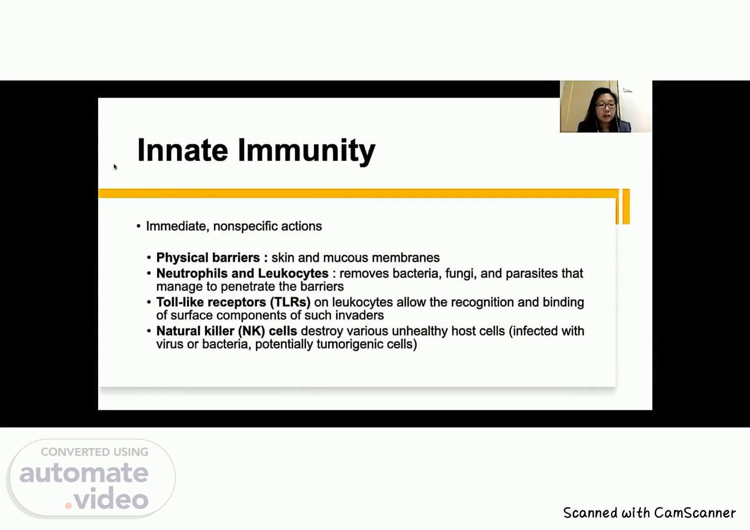
. . . Innate Immunity • Immediate, nonspecific actions • Physical barriers : skin and mucous membranes • Neutrophils and Leukocytes : removes bacteria, fungi, and parasites that manage to penetrate the barriers • Toll-like receptors (TLRs) on leukocytes allow the recognition and binding of surface components of such invaders • Natural killer (NK) cells destroy various unhealthy host cells (infected with virus or bacteria, potentially tumorigenic cells).
Scene 2 (18s)
. . . 13:13 Hisco 01:37 .ill Antimicrobial Chemicals • Hydrochloric acid (HCI) and organic acids • Lower the pH locally to either kill entering microorganisms directly or inhibit their growth. • Defensins • Produced by neutrophils and various epithelial cells that kill bacteria by disrupting the cell walls. • Lysozyme • enzyme made by neutrophils and cells of epithelial barriers, which hydrolyzes bacterial cell wall components, killing those cells. Speed.
Scene 3 (37s)
. . . Antimicrobial Chemicals • Hydrochloric acid (HCI) and organic acids • Lower the pH locally to either kill entering microorganisms directly or inhibit their growth. • Defensins • Produced by neutrophils and various epithelial cells that kill bacteria by disrupting the cell walls. • Lysozyme • enzyme made by neutrophils and cells of epithelial barriers, which hydrolyzes bacterial cell wall components, killing those cells..
Scene 4 (55s)
. . . Antimicrobial Chemicals • Complement a system of proteins in blood plasma, mucus, and macrophages that react with bacterial surface components to aid removal of bacteria. • Interferons • paracrine factors from leukocytes and virus-infected cells that signal NK cells to kill such cells and adjacent cells to resist viral infection..
Scene 5 (1m 11s)
. . . Adaptive Immunity Four primary characteristics: .immunological memory .immunological specificity .immunological diversity .capability to differentiate between self and nonself..
Scene 6 (1m 22s)
. . . Adaptive immunity • B and T lymphocytes • Activated against specific invaders by being presented with specific molecules from those cells by APCs, which are usually derived from monocytes. • Aimed at specific microbial invaders and involve production of memory lymphocytes.
Scene 7 (1m 35s)
. . . CYTOKINES • How the immune system communicates with each other • Involved in both innate and adaptive immunity • Diverse group of peptides and glycoproteins.
Scene 8 (1m 46s)
. . . Cytokines: Major responses induced in target cells • Chemotaxis: directs cell movements toward and cell accumulation at sites of inflammation • Cytokines producing this effect are also called chemokines • Interleukins (a group of cytokines) that stimulate or suppress lymphocyte activities in adaptive immunity. • Stimulates phagocytosis or directs cell killing by innate immune cells.
Scene 9 (2m 2s)
. . . ANTIGENS • A molecule recognized by cells of the adaptive immune system • Soluble molecules (proteins or polysaccharides or molecules that are still components of intact cells (bacteria, protozoa, or tumor cells . • Epitopes - small molecular domains of antigen that Immune cells recognize and react to • Immune response to antigens may be cellular (lymphocytes), humoral (antibodies), or both.
Scene 10 (2m 19s)
. . . Antibody ANTIGEN VS ANTIBODY • A glycoprotein of the immunoglobulin family that interacts specifically with an antigenic determinant. • Secreted by plasma cells dreemÆime,corn.
Scene 11 (2m 29s)
. . . Antibody FIGURE 14—2 Basicstructureof•n Immunoglobulin (antibody). Antiwn-bhdng site Disuffde sae Variaue regim Cmstant regim regbn (Fab) Cel biMirv Heavy • Two identical light chains • Two identical heavy chains bound by disulfide bonds TWO light chains and two heavy chains form an antibody ecule (•monomer•). The chains are linked by disulfide bonds. The variable portions (Fab) near the amino end of the Eght and.
Scene 12 (2m 45s)
. . . FIGURE 14—2 Basic structure of an Antibody • Heavy-chain molecules- • Recognized by cell surface receptors on basophils and mast cells, localizing these antibodies to the surface of these cells. Antigen-binding site Disulfide Hinge region Variable Constant Antigen-binfig site Antiwi•binding (Fab) porion Light chain Cell (Fc) Heavy chain Two light chains and two heavy chains form an antibody mol- ("monomer). The chains are linked by disulfide bonds. The variable portions (Fab) near the amino end Of the light and.
Scene 13 (3m 4s)
. . . Antibody • Variable region : • Make up an antibody's antigen- binding site. • DNA sequences coding for these regions undergo recombination and rearrangement after B lymphocytes are activated against a specific antigen FIGURE 14—2 Basic structure ofan immunoglobulin Arigen-bin&rv Variauo Cmstmt chain Cell YMing Heavy cruin TWO chains and two heavy chains form an mol- Cmonomer•). The chains are linked by disulfide The variable portions (Fab) near the amino end Of the light and heavy chains bind the antigen. The constant region (or Fc) of the molecule m" bind to surface receptors of several cen types..
Scene 14 (3m 27s)
. . . Classes of Antibodies Important features of the antibody classes in humans. TABLE 14—2 Structure Antibody in the plasma Known furxdons IgG Monomer Fetal Circulation in pregnant women neutralizes antÉens passive Immunity IgM Pentamer 596-10% B lymphocyte surface (as a monomer) First in initial immune respnsg activates compkrnmt IgA secretory component Dimer with J chain and secretory component Secretions (saliva, milk. tears, etc) Protects mucosae lgD Monomer Surface Of B lymphxytes Antigen receptor triggering initial B cell activation lgE Monomer 0.002% Bound to the surface of mast cells and basophils Destroys parasitic worms and parti- cipates in allergies.
Scene 15 (3m 45s)
. . . Binding of antigen-binding site of an antibody with antigen causes: Neutralization Antibody covers biologically active portion Of microbe or toxin. Agglutination Antibody cross-links cells (eg, bacteria). forming a •clump." Antigen Bacteria Preclpltatlon Antibody cross-links circulating particles (eg, toxins), forming an insoluble antigen-antibody complex. Soluble particles Antigen-antibody complex Antibody.
Scene 16 (4m 0s)
. . . Exposed Fc portion following antigen binding by antibody promotes: Complement fixation Fc region of antibody binds complement proteins; complement is activated. Bacterium Fc region of antibody Opsonlzatlon Fc region of antibody binds to receptors of phagocytic cells, triggering phagocytosis. Bacterium Fc region of antibody Receptor for Fc region of antibody Activation of NK cells Fc region 01 antibody binds to an NK cell, triggering release of wtotoxic chernicals. PerforW Virus-infected cell Receptor for Fc region Of antibody Antibody Shown here are important mechanisms by which the most com- mon antibodies act in immunity. (a) Specific binding of antigens can neutralize or precipitate antigens, or cause microorganisms bearing the antigens to dump (agglutinate) for easier removal. (b) Complement proteins and surface receptors on many leuko- cytes bind the Fc portions of antibodies attached to cell-surface antigens, producing active complement. more efficient phagocy- tosis (opsonizadon), and NK-celI activation..
Scene 17 (4m 33s)
. . . Major histocompatibility complex (MHC) • MHC class I and class II. • First recognized by their roles in the immune rejection of grafted tissue or organs. • Proteins of both classes: human leukocyte antigens (HLAs) • T lymphocytes are specialized to recognize both classes of MHC proteins and the antigens they present.
Scene 18 (4m 48s)
. . . CLINICAL CORRELATION • Autografts - when the donor and the host are the same individual (burn patient) • Isografts are those involving identical twins. • Homografts (or allografts), which involve two related or unrelated individuals • Cyclosporin drug: inhibit activation of cytotoxic T cells; allowed the more widespread use of allografts or even xenografts (animal donor).
Scene 19 (5m 5s)
. . . CELLS OF ADAPTIVE IMMUNITY • Antigen-Presenting Cells • all types of macrophages and specialized dendritic cells in lymphoid organs. • active endocytotic system and expression of MHC class II molecules for presenting peptides of exogenous antigens.
Scene 20 (5m 17s)
. . . CELLS OF ADAPTIVE IMMUNITY TABLE 14-3 • Lymphocyte s • Adaptive immunity • Band T • Do not stay long in the lymphoid organs • Continuously recirculate through the body in connective tissues, blood, and lymph. Approximate percentages o and T cells in lymphoid orga T Lymphocytes hoid Organ %) Thymus Bone marrow Spleen Lymph nodes 100 10 45 60 70 B Lymph 90 55 40 30.
Scene 21 (5m 32s)
. . . T Lymphocytes T lymphocytes • Helper T cells - assist other lymphocytes by secreting immune chemicals called cytokines Cytotoxic T cells - recognize antigenically different cells such as virus-infected cells, foreign cells, or malignant cells and destroy them. • Memory T cells - long-living progeny of T cells; respond rapidly to the same antigens in the body and stimulate immediate production of cytotoxic T cells. • Suppressor T cells - decrease or inhibit the functions of helper T cells and cytotoxic T cells; modulate the immune response..
Scene 22 (5m 53s)
. . . B Lymphocytes • Plasma cells • From differentiated B Lymphocytes • secretes antibodies that will bind the same epitope recognized by the activated B cell. • Humoral immunity • As with activated T cells, some of the newly formed B cells remain as long-lived memory B cells • Formation of long-lived memory lymphocytes is a key feature of adaptive immunity, which allows a very rapid response upon subsequent exposure to the same antigen..
Scene 23 (6m 12s)
. . . Lymphoid System •Collects excess interstitial fluid into lymphatic capillaries •Transports absorbed lipids from the small intestine • Responds immunologically to invading foreign substances. •Main function - protect the organism against invading pathogens or antigens (bacteria, parasites, and viruses). •Wide distribution in the body. eo uNrVERStry OF PERPETUAL HELP SYSTEM DALIA or.
Scene 24 (6m 28s)
. . . Lymphoid System • The lymphoid system includes am cells, tissues, and organs in the body that contain aggregates of immune cells called lymphocytes. • Cells of the immune system, especially lymphocytes, are distributed throughout the body in the loose connective tissue of digestive, respiratory, and reproductive systems, or as encapsulated individual lymphoid organs. • Major lymphoid organs: • lymph nodes, tonsils, thymus, and spleen. • the bone marrow produces lymphocytes, and is considered a lymphoid organ UNIVERSITY OF PERPETUAL HELP SYSTEM DALTA SCHOOL or.
Scene 25 (6m 52s)
. . . UNIVERSITY OF PERPETUAL HELP SYSTEM DALIA or.
Scene 26 (6m 59s)
. . . Lymph Node • Bean-shaped, encapsulated structures • NIO mm by 2.5 cm in size • distributed throughout the body along the lymphatic vessels • 400 to 450 lymph nodes : in the axillae (armpits) and groin, along the major vessels of the neck, and in the thorax and abdomen, and especially in the visceral mesenteries eo UNIVERSITY OF PERPETUAL HELP SYSTEM DALTA.
Scene 27 (7m 17s)
. . . Lymph Node • Convex surface where afferent lymphatics enter • Hilum - Concave depression where an efferent lymphatic leaves where an artery, vein, and nerve penetrate the organ • Capsule - dense connective tissue capsule. UNIVERSITY OF PERPETuAL HELP SYSTEM DALTA.
Scene 28 (7m 31s)
. . . Lymph Node Dense masses of lymphocyte aggregations intermixed with dilated lymphatic sinuses that contain lymph and are supported by a framework of fine reticular fibers. Outer cortex Inner medulla Surrounded by a pericapsular adipose tissue A dense connective tissue capsule surrounds the lymph node. Stan: UNIVERSITY OF PERPETUAL HELP SYSTEM DALIA SCHOOL or.
Scene 29 (7m 46s)
. . . LymNtÉ Anormt in T as as h tho Stan:.
Scene 30 (7m 53s)
. . . Lymph Node Connective tissue trabeculae extend into the node Afferent lymphatic vessels with valves course in the connective tissue capsule of the lymph node and penetrate the capsule to enter a narrow space called the subcapsular sinus The sinuses (cortical sinuses) extend along the trabeculae to pass into the medullary sinuses • Stan: UNIVERSITY OF PERPETUAL HELP SYSTEM DALIA.
Scene 31 (8m 9s)
. . . ox Stan: uNtVERSITY OF PERPETUAL HELP SYSTEM DALTA SCHOOL cr Lymph Node The cortex of the lymph node contains numerous lymphocyte aggregations called lymphatic nodules. Germinal centers represent the active sites of lymphocyte proliferation..
Scene 32 (8m 21s)
. . . Lymph Node Lymphocytes are arranged as irregular cords of lymphatic tissue called medullary cords. Contain macrophages, plasma cells, and small lymphocytes. Medullary sinuses drain the lymph from the cortical region of the lymph node and course between the medullary cords toward the hilus of the organ. • Stan: VNNERSITY OF PERPETUAL HELP SYSTEM DALTA SCHOOL cr.
Scene 33 (8m 38s)
. . . Lymph Node Lymphatic nodules exhibit a central, light- staining germinal center surrounded by a deeper-staining peripheral portion of the nodule. In germinal centers, the cells are more loosely aggregated and the developing lymphocytes have larger and lighter-staining nuclei with more cytoplasm. 92 Lymph node: capsule. Stain: and maplifkatlm. UNIVERSITY OF PERPETUAL HELP SYSTEM DALTA.
Scene 34 (8m 55s)
. . . Lymph Node IS ea.r.aggnex: thymus-dependent zone and is primarily occupied by I_ggLls: Transition area from the lymphatic nodules to the medullary cords of the lymph node medulla. Medulla: anastomosing cords of lymphatic tissue/medullary cords, interspersed with medullary sinuses that drain the lymph from the node into the efferent lymphatic vessels located at the hilus •O t.fr•.:;.. :..Ö.• FIGURE 92 Lymph capsule. Stain: and UNIVERSITY OF PERPETUAL HELP SYSTEM DALIA.
Scene 35 (9m 17s)
. . . .
Scene 36 (9m 23s)
. . . Ch"d Thymus Gland • Soft, lobulated lymphoepithelial organ located in the upper anterior mediastinum and lower part of the neck. • Most active during childhood, then undergoes slow involution • Filled with adipose tissue in adults (C) Of th•nu..
Scene 37 (9m 36s)
. . . Thymus Gland Surrounded by a connective tissue capsule Cortex Dark-staining with an extensive network of interconnecting spaces Colonized by immature lymphocytes that migrate here from hemopoietic tissues in the developing individual to undergo maturation and differentiation. Medulla lighter-staining; epithelial cells form a coarser framework that contains fewer lymphocytes and whorls of epithelial cells that combine to form thymic (Hassall's) corpuscles..
Scene 38 (9m 53s)
. . . us Gland 10 C.aßdat veül Hagan (thymk) • plat-uf tnanmamir qain• hematnrvlin and msin I.
Scene 39 (10m 2s)
. . . Thymus Gland FICURE9S • Calex of a Stain: hematoxylin emin.
Scene 40 (10m 10s)
. . . Thymus Gland Hassall's corpuscles are oval structures consisting of round or spherical aggregations (whorls) of flattened epithelial cells. Exhibit calcification or degeneration centers that stain pink or eosinophilic. The functional significance of these corpuscles remains unknown. S Cain 10 FIGURE 9.8 • Stain: hematoxym.
Scene 41 (10m 24s)
. . . Spleen • Largest lymphoid organ of the body • Functions: • filter blood • phagocytose senescent RBC and invading microorganisms • supply immunocompetent T and B lymphocytes, • manufacture antibodies. VNtVERStry OF PERPETUAL HELP SYSTEM T Splenic vein Splenic artery.
Scene 42 (10m 36s)
. . . Spleen • Not divided into cortical and medullary regions • Not supplied by afferent lymphatic vessels. • Blood vessels enter and leave the spleen at its hilum and travel within the parenchyma via trabeculae derived from its connective tissue capsule Splene UNIVERSITY OF PERPETUAL HELP SYSTEM DALTA SCHOOL or "CURE qw • Spiem "an: arw mam«atøn.
Scene 43 (10m 52s)
. . . Spleen • Large trabeculae originate at the hilum and carry branches of the splenic artery, vein, lymphati&, and nerves into the splenic pulp. UNNERStry OF PERPETUAL HELP SYSTEM DALTA • "in: herrutoxMh mam«atøn.
Scene 44 (11m 5s)
. . . Spleen • Composed of lymphoid tissue arranged in a specific fashion, either as: • Periarterial lymphatic sheaths (PALS) - T lymphocytes or • Lymphoid nodules - B lymphocytes UNIVERSITY OF PERPETUAL HELP SYSTEM DALTA or.
Scene 45 (11m 16s)
. . . Spleen Red Pulp • Red Pulp Consists of pulp cords (of Billroth) interposed between a spongy network of sinusoids lined by unusual elongated endothelial cells displaying large intercellular spaces UNIVERSITY OF PERPETUAL HELP SYSTEM DALIA.
Scene 46 (11m 28s)
. . . Spleen • Red pulp • Supported by a thick, discontinuous, hoop-like basement membrane. Reticular cells and reticular fibers associated With these sinusoids extend into the pulp cords to contribute to the cell population that consists of macrophages, plasma cells, and extravasated blood cells. UNIVERSITY OF PERPETUAL HELP SYSTEM DALIA.
Scene 47 (11m 44s)
. . . Spleen • Mergirl?' Zone Region of smaller sinusoids forming the interface between the white and red pulps Capillaries arising from the central arteries deliver their blood to sinusoids of the marginal zone Rich in arterial vessels and phagocytic macrophages. APCs of the marginal zone monitor this blood for the presence of antigens and foreign substances. UNIVERSITY OF PERPETUAL HELP SYSTEM DALIA 22 (PALS) B Roa.
Scene 48 (12m 1s)
. . . Spleen • The splenic artery entering at the hilum is distributed to the interior of the organ via trabeculae as trabecular arteries. FIGURE 14-21 Spleen. 22 in the •plnn. (PALS) B Rod UNNERSITY OF PERPETUAL HELP SYSTEM DALTA.
Scene 49 (12m 14s)
. . . Spleen Cenbal arteries enter the red pulp by 22 losing their PALS and subdivide into numerous small, straight vessels known as penicillar arteries. UNIVERSITY OF PERPETUAL HELP SYSTEM DALIA in th• •plnn. (PALS) B Red.
Scene 50 (12m 26s)
. . . Spleen • • Penicillar arteries possess three regions: pulp arterioles, sheathed arterioles, and terminal arterial capillaries. Whether these terminal arterial capillaries drain directly into the sinusoids (closed circulation) or ter- minate as open-ended vessels in the pulp cords (open circulation) has not been determined conclusively; in humans, the open circulation is believed to predominate. 22 in the spl«n. (PALS) B Red eo UNIVERSITY OF PERPETUAL HELP SYSTEM DALIA or.