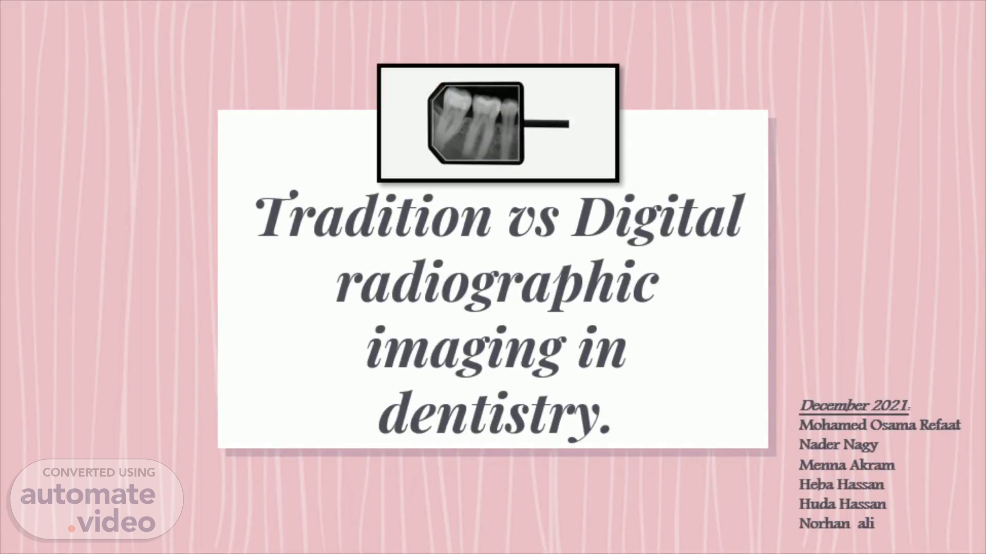
Tradition vs Digital radiographic imaging in dentistry.
Scene 1 (0s)
Tradition vs Digital radiographic imaging in dentistry..
Scene 2 (11s)
The importance of dental X-rays. Detecting the invisibile . X-rays facilitate viewing structures that are clinically undetectable such as bone density, tooth apex and lamina dura; thus making the discovery of small lesions a possibility..
Scene 3 (1m 38s)
Types of dental x rays. 3. Bite wing Show upper and lower crowns and parts of the roots to about the level of the supporting bone To detect proximal caries -changes in bone density caused by gum disease -proper fit of a crown (or cast restoration) and the marginal integrity of fillings..
Scene 4 (6m 10s)
Methods of capturing dental X-rays. Are either by using a traditional x ray film Or using a digital sensor Being different in their structural formation, the x-ray sensors and films differ in their radiographic capabilities and imaging qualities..
Scene 5 (7m 0s)
Traditional films. X-ray film displays the radiographic image and consists of emulsion (single or double) of silver halide (silver bromide ( AgBr ) is most common) which when exposed to light, produces a silver ion (Ag+) and an electron. The electrons get attached to the sensitivity specks and attract the silver ion. Subsequently, the silver ions attach and clumps of metallic silver (black) are formed. Thus, film-based images are described as analogue images..
Scene 6 (7m 37s)
Traditional film compositon. 6. The intraoral x-ray films - Dental Care.
Scene 7 (7m 56s)
Digital sensors. The image receptor used instead of the xray film is called the sensor or the image plate(IP). The idea depends on converting the xray energy reaching the image receptor into electric energy in the form of electronic signals to be analyzed and digitalized by the computer which in turn generates what is so called the ‘Digital image’..
Scene 8 (8m 25s)
Digital sensor compositon. 8. • posed • static which cause powet« erasing the plate it.
Scene 9 (8m 49s)
Image Processing. In traditional films: Developing: will take the bromide part of all crystals that have exposed silver halide molecules Rinsing : the film is rinsed in water for 10 -20 s with continuous gentle agitation before they are placed in fixer Fixation : to fix the precipitated black silver in place and to remove the undeveloped crystals and to harden the film emulsion Washing : proper time 20:30 minutes, improper washing dicolors and causes stains on the film But remember not to overload your slides with content Drying: to facilitate film handling.
Scene 10 (9m 40s)
Image Processing. In Digital sensors: Uses sensor of solid-state, and information is presented and stored as an image using a computer. There are two types: Charged-coupled device (CCD) Photostimulable phosphor (PSP).
Scene 11 (9m 53s)
(a) Electric charge is formed by X-ray exposure. The charge is temporarily stored in each vertical shift register. (b) Each charge moves to the adjacent register by applying a voltage. The charge reaching the end of a vertical shift register moves to a horizontal shift register. (c) and (d) Each charge moves sequentially.
Scene 12 (10m 20s)
(a) The crystal of BaFBr:Eu is exposed. (b) Ionization of Eu occurs, and Eu2+ turns into Eu3+. The ionizing electron is trapped in BaFBr . (c) In reading process, the exposed crystal is irradiated by laser. (d) The laser irradiation affects the electron trap, and the electron recombines with Eu3+. Therefore Eu3+ returns to Eu2+, and the energy difference produces fluorescence. The fluorescence is measured by detector.
Scene 13 (11m 2s)
1) Instant viewing of images: the film is immediately processed and available to view, whereas conventional film takes time to be developed 2) Less radiation needed to produce the same quality image as film (digital X-rays gives 70% less exposure to radiation than conventional X-rays). 3) Eliminates the need for darkroom and chemicals, and doesn’t generate hazardous waste..
Scene 14 (11m 46s)
4) Improved grayscale may aid in diagnosis 5) Image adjustment for better visualization: the image can be enlarged, the contrast and brightness can be changed, and the image can be inverted or rotated. 6) Electronic records that can be transferred to the patient’s insurance company or specialist 7) No need for film duplication because images can be reprinted without loss of image quality 8) Don’t degrade overtime 9) Less physical storage space required 10) Provides extremely efficient progress evaluation during endodontic and oral surgery procedures..
Scene 15 (13m 10s)
Disadvantage of digital radiograph over conventional radiograph.
Scene 16 (14m 29s)
Thank You.. 16.