ANATOMY TO KNOW HEART
Scene 1 (0s)
ANATOMY TO KNOW HEART. S IMPLE I MPORTANT P RACTICAL.
Scene 2 (7s)
CARDIOLOGY The scientific study of the normal heart and the diseases associated with it CARDIO LOGY HEART STUDY OF.
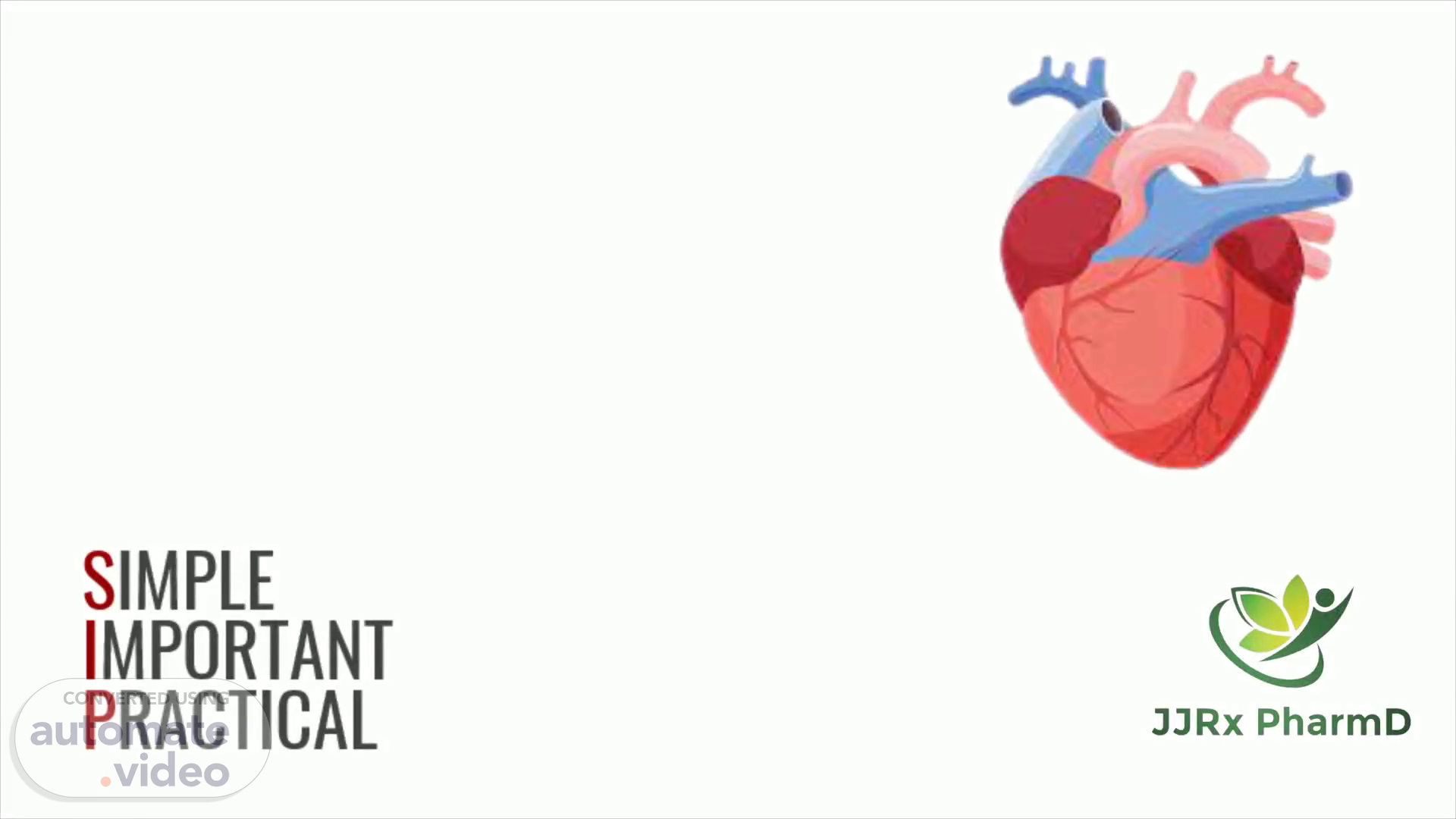
Scene 4 (23s)
DEFINITION An organ that pumps blood throughout the body via the vessels of the circulatory system, supplying oxygen and nutrients to the tissues and removing carbon dioxide and other wastes..
Scene 5 (35s)
STRUCTURAL CHARACTERISTICS Shape: Hollow cone shaped Size: Length- 12cm Width- 9cm Thickness- 6 cm Weight: Males- approx. 300g Females- approx. 250g.
Scene 6 (47s)
LOCATION Lies in the space between the lungs called Mediastinum which is- In the middle of the chest, Just behind the sternum, Above the diaphragm. 2/3rd of the Heart → lies to the left of sternum. Remaining 1/3rd of the heart → lies to the right of sternum..
Scene 7 (1m 3s)
PARTS.
Scene 9 (1m 15s)
M ade up of four chambers: Two upper chambers - the left & right atrium Two lower chambers - the left & right ventricles Apex : Pointed, formed by the tip of the lower ventricle & r est on the diaphragm at the level of 5th intercostal space. Base: opposite of the apex, formed by atria mostly left atrium w hich extended to the level of 2nd rib..
Scene 10 (1m 33s)
Has different surfaces & borders. Anterior surface: deep to the sternum & ribs. Inferior surface: between the apex & right surface & rest mostly on the diaphragm. Right surface: faces right lung & extends from the i nferior surface to the base. Left surface: faces the left lung & extends from the b ase to the apex..
Scene 11 (1m 50s)
PERICARDIUM Membrane that surrounds & protects the heart. Confines the heart to its position in the mediastinum. Consists of two main parts: i . Fibrous pericardium i i. Serous pericardium.
Scene 12 (2m 2s)
PERICARDIUM i. Fibrous pericardium Composed of tough, inelastic, dense irregular connective tissue. Resembles a bag that rests on & attaches to the d iaphragm. Prevents overstretching of the heart, provides protection & anchors the heart in the mediastinum..
Scene 13 (2m 16s)
PERICARDIUM ii. Serous pericardium Thinner, more delicate membrane that forms a double a round the heart Outer parietal layer- fused to the fibrous pericardium Inner visceral layer- epicardium (one of the layers of the h eart wall & adheres tightly to the surface of the heart).
Scene 14 (2m 30s)
PERICARDIUM Pericardial fluid:- Between the parietal & visceral layers of the serous pericardium - thin film of lubricating serous fluid. This slippery secretion of the pericardial cells, known as pericardial fluid Pericardial cavity:- Space that contain few milliliters of pericardial fluid..
Scene 15 (2m 44s)
LAYERS OF THE HEART WALL Consists of three layers - i . Epicardium i i. Myocardium i ii. Endocardium.
Scene 16 (2m 53s)
LAYERS OF THE HEART WALL i. Epicardium Outermost transparent layer of heart Composed of two tissues- visceral layer of serous p ericardium & mesothelium. Mesothelium- variable layer of delicare fibroelastic & a dipose tissue. Imparts a smooth, slippery texture to the outermost layer..
Scene 17 (3m 7s)
LAYERS OF THE HEART WALL ii. Myocardium Second layer of the heart wall Responsible for pumping action of heart. Composed of cardiac muscle Makes up approx 95% of the heart..
Scene 18 (3m 19s)
LAYERS OF THE HEART WALL iii. Endocardium Thin innermost layer of the heart wall. Made up of glistening sheet of endothelium. Covers the heart chambers, heart valves & blood vessels that enter, leave the heart. Allows smooth flow of blood in the heart.
Scene 19 (3m 34s)
CHAMB ERS OF THE HEART Has four chambers i. Right atrium ii. Left atrium iii. Right ventricle iv. Left ventricle.
Scene 20 (3m 44s)
CHAMBERS OF THE HEART Atria :- Upper two chambers that receive blood from different parts of the body Wrinkled pouch like structure- auricle Auricles increases the blood holding capacity of each atrium 2 thin walled atria are separated by a thin portion- interatrial septum..
Scene 21 (3m 59s)
CHAMBERS OF THE HEART i. Right atrium Forms the right surface of the heart Receives deoxygenated blood from three veins- Superior vena cava Inferior vena cava Coronary sinus Thickness: 2-3mm.
Scene 22 (4m 11s)
CHAMBERS OF THE HEART i. Right atrium Superior vena cava bring blood from arms, head & neck. Inferior vena cava bring blood from abdomen & legs. Coronary sinus brings blood from the vessels that supply blood to the heart walls.
Scene 23 (4m 24s)
CHAMBERS OF THE HEART ii. Left atrium Thickness: 2-3 mm Receives oxygenated blood from the lungs via 4 pulmonary veins Blood passes from the left atrium via bicuspid valve..
Scene 24 (4m 36s)
CHAMBERS OF THE HEART Ventricles :- Two chambers that pump blood to distinct parts of the body. Receives blood from atria On anterior portion, two ventricles are separated externally from each other by anterior interventricular sulcus & posteriorly by posterior interventricular sulcus ..
Scene 25 (4m 51s)
CHAMBERS OF THE HEART iii. Right ventricle Thickness: 4-5 mm. Forms most of the anterior surface of the heart. Receives deoxygenated blood from the right atrium via tricuspid valve..
Scene 26 (5m 3s)
CHAMBERS OF THE HEART iv. Left ventricle Forms the apex of the heart. Thickness: 4-5 mm. Receives blood from the left atrium via bicuspid valve. Blood from the left ventricle is pumped at a high pressure via aortic valve into the ascending aorta..
Scene 27 (5m 18s)
CARDIAC MUSCLE Important tissue of the heart Acquires nutrition in the form of ATP, calcium ions, oxygen, p otassium ions & sodium ions to perform its function Types:- Contractile Nodal Conducting.
Scene 28 (5m 30s)
CARDIAC MUSCLE Properties of cardiac muscle tissues:- i . Excitability or Irritability i i. C ontractility iii. Automaticity & Conductivity i v. Rhythmicity v . Refractory period v i. Post extrasystolic potentiation vii. Tonicity.
Scene 29 (5m 43s)
THANK YOU.